
Dr. Erik Sontheimer – Innovation Is in Our RNA
DNA, as the molecular blueprint for life on earth, has long held a special place in scientific discourse and popular culture. DNA also has a lesser-known sister molecule, called RNA, which transfers specific instructions from DNA to produce proteins. RNA is now recognized as having a diverse range of previously unimaginable roles beyond protein production. Dr. Erik Sontheimer of the RNA Therapeutics Institute, UMass Medical School, has devoted his career to understanding this RNA world present in every cell.
Protein molecules, with their diverse range of functional, structural, catalytic and regulatory roles, ultimately dominate an organism’s set of observable traits, its phenotype. The information for creating proteins is stored in DNA, in units known as genes. DNA is a hereditary molecule, a set of instructions that are passed down through generations.
The science of molecular biology is concerned with elucidating the intricate mechanisms behind the information flow from DNA to proteins – from genotype to phenotype. Between DNA and proteins, information is transmitted by RNA molecules – or more specifically, messenger RNA (mRNA). During transcription, the information encoded in a gene is transcribed onto a corresponding newly-synthesized mRNA molecule. This mRNA molecule then diffuses to a ribosome – the cellular protein factory – where amino acids (of which there are 20 types) are joined together into a specific assortment to form a chain based on the instructions given by the mRNA molecule. This process is known as translation. The chain then folds into a specific shape to form a protein that goes on to carry out a particular function in the cell.
However, the full biological scope of RNA extends way beyond its roles in protein production. In the past few decades, molecular biologists have come to realize that there is a diverse range of RNA molecules in every cell that do not encode proteins (non-coding RNAs). These RNAs have various biochemical functions, many of which are not yet fully known. In fact, we are barely scratching the surface of this mysterious RNA world.
DNA and RNA are both nucleic acids – linear molecules comprising strands of nucleotide units. These nucleotides each contain one of four different bases – A, T (or U in RNA), G and C – and are arranged in various combinations, with groups of three sequential bases – known as codons – coding for each amino acid. This code is transferred from DNA, to RNA, and then to proteins during gene expression, and is universally present in all living organisms – both prokaryotes (organisms with simple cells such as bacteria), and eukaryotes (organisms with complex cells such as plants and animals). This genetic code, transferred from DNA to RNA to proteins, may be likened to text – with each codon of three bases and corresponding amino acid as one letter, combining in specific combinations to form words known as proteins – which write an organism’s phenotype.
E-book
Reference
https://doi.org/10.26320/SCIENTIA66
Share
Revelations in RNA Biology
Dr. Sontheimer’s research into RNA has been extensive as well as insightful. When he started as a grad student at Yale in 1987, it was a time of great progress in RNA science. The previous 10 years had seen several fundamental discoveries within the RNA space. “It looked to me like the field was hitting its stride, with lots of room for more big discoveries,” Dr. Sontheimer reflects. “This was particularly easy to recognize at Yale, which was (and remains) a leading center of RNA research.”
In eukaryotes, gene expression is fairly complicated. Eukaryotic genes contain series of protein-coding DNA sequences, known as exons interdispersed with long stretches of DNA that do not code for protein, known as introns, both of which are transcribed into an mRNA molecule. The roles of these non-coding introns are still mysterious, but they must be removed (spliced out) from the mRNA molecule before proteins are created from the instructions it carries. This is mediated by the spliceosome – a massive enzyme composed of over 100 proteins and five small nuclear RNAs (snRNAs) that removes (splices) introns from the mRNA molecule, and can join the cut ends back together. Like a pair of scissors, the spliceosome cuts out the introns, leaving only protein-coding sequences in the mRNA.
Dr. Sontheimer’s grad school studies focused on investigating the assembly, structure and splicing mechanism of the spliceosome. After gaining his PhD, Dr. Sontheimer worked as a postdoctoral fellow at the University of Chicago, where he engineered changes in long mRNAs, and found metal atoms in the active site of the spliceosome enzyme, providing the first insights into the chemical mechanism of mRNA splicing. Dr. Sontheimer’s later research trajectory reflected the prevailing currents of the RNA field. “The field’s potential turned out to be even greater than I suspected,” he says. In 1999, he started working as a researcher at Northwestern University, where he studied RNA silencing complexes.
In eukaryotes, RNA interference (RNAi) – a process by which a transcribed mRNA is degraded or prevented from being translated to new proteins – plays an important role in regulating gene expression. RNAi is mediated by tiny non-coding RNA molecules, particularly small interfering RNAs (siRNAs) and microRNAs (miRNAs). In 2002, Dr. Sontheimer began collaborating with Dr. Rich Carthew (also at Northwestern) to elucidate the mechanisms of RNAi, employing the fruit fly as a model organism. siRNAs act through a large molecule known as the siRNA-induced silencing complex (siRISC), which targets and destroys mRNA molecules, preventing them from being translated into proteins. One of the team’s most surprising discoveries is that a protein called Dicer2 is involved in the assembly of the siRISC complex.
In 2007, an RNA-based immune system was discovered in bacteria, known as CRISPR. This discovery soon started a revolution when CRISPR began to be repurposed as a DNA-editing toolkit. Dr. Sontheimer was one of the earliest proponents of CRISPR biotechnology, realizing that it could be a democratizing tool for genetic programming. His group has since been pursuing this research theme, first at Northwestern, and more recently in the RNA Therapeutics Institute (RTI) at the University of Massachusetts Medical School, where he moved in 2014. In line with the RTI’s ethos of applying RNA biology to preventing and curing diseases, his current work with the NmeCas9-based CRISPR system is geared towards therapeutic gene editing. But what is CRISPR, anyway? And why does it have such great biotechnological potential?
Engineering CRISPR Systems
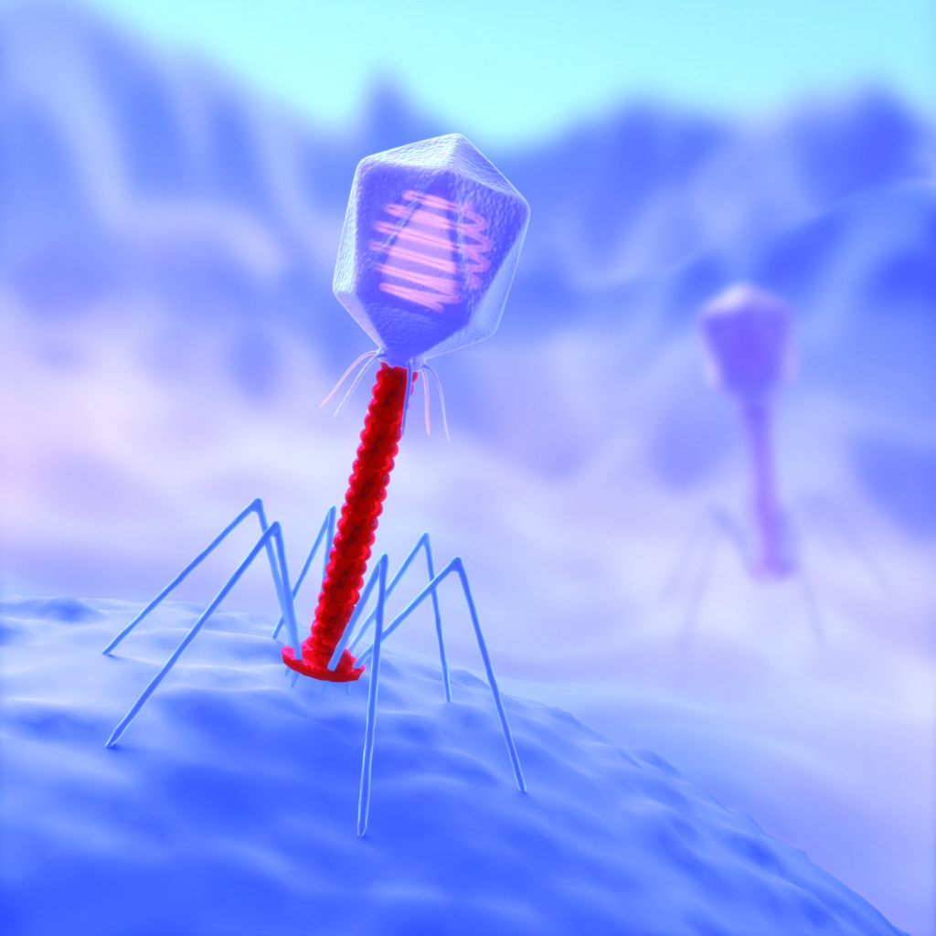
Bacteria are under constant attack from viruses known as phages, which hijack the bacterial cell by inserting their own genes that express viral proteins, leading to the production of more phages in a perpetual cycle. Fortunately, bacteria have evolved several immune responses against viral attack – including the CRISPR-Cas system, known informally as CRISPR. CRISPR actually refers to “clustered, regularly interspaced, short palindromic repeats”, which are specialized sequences within bacterial DNA.
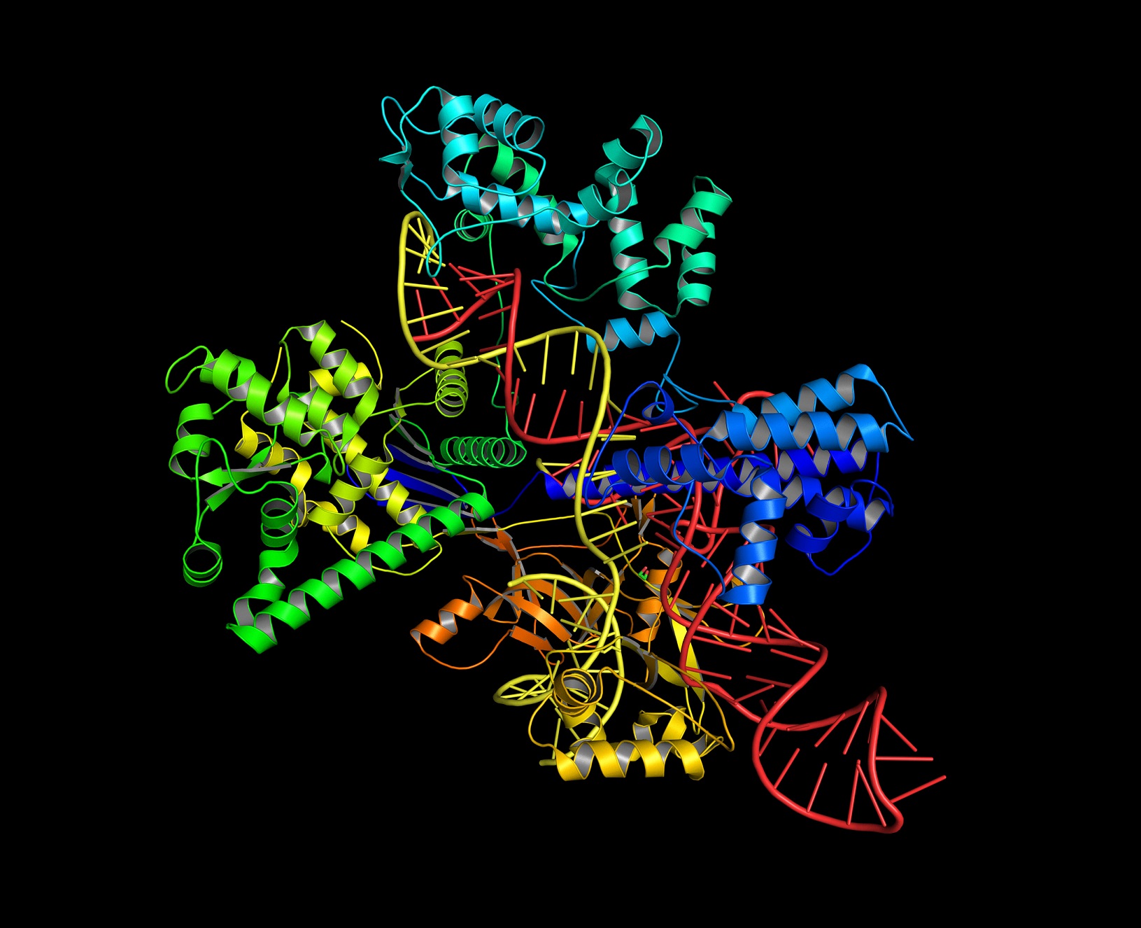
In the late 1980s, biologists first reported these strange arrays of DNA repeats in bacterial genomes. There were several proposed hypotheses for their existence, though no one really knew their purpose, or their eventual significance. Only in the mid-2000s was the purpose of CRISPR determined – it turned out to be an adaptive immune system to protect the bacterial genome against viral attack. When a bacterial cell becomes infected with a virus, a piece of viral DNA, known as a spacer, is integrated into the CRISPR sequence to vaccinate the cell. Spacers are transcribed into small RNA molecules – crRNAs (CRISPR RNAs) – that associate with Cas proteins, such as Cas9 to create crRNA-Cas complexes. Cas proteins include enzymes that cut DNA strands at specific locations. The crRNA-Cas complex is like a guided missile, with the crRNA guiding the Cas protein to specific sections of the viral genome and the Cas protein chewing up the viral DNA (and in some cases, viral RNA).
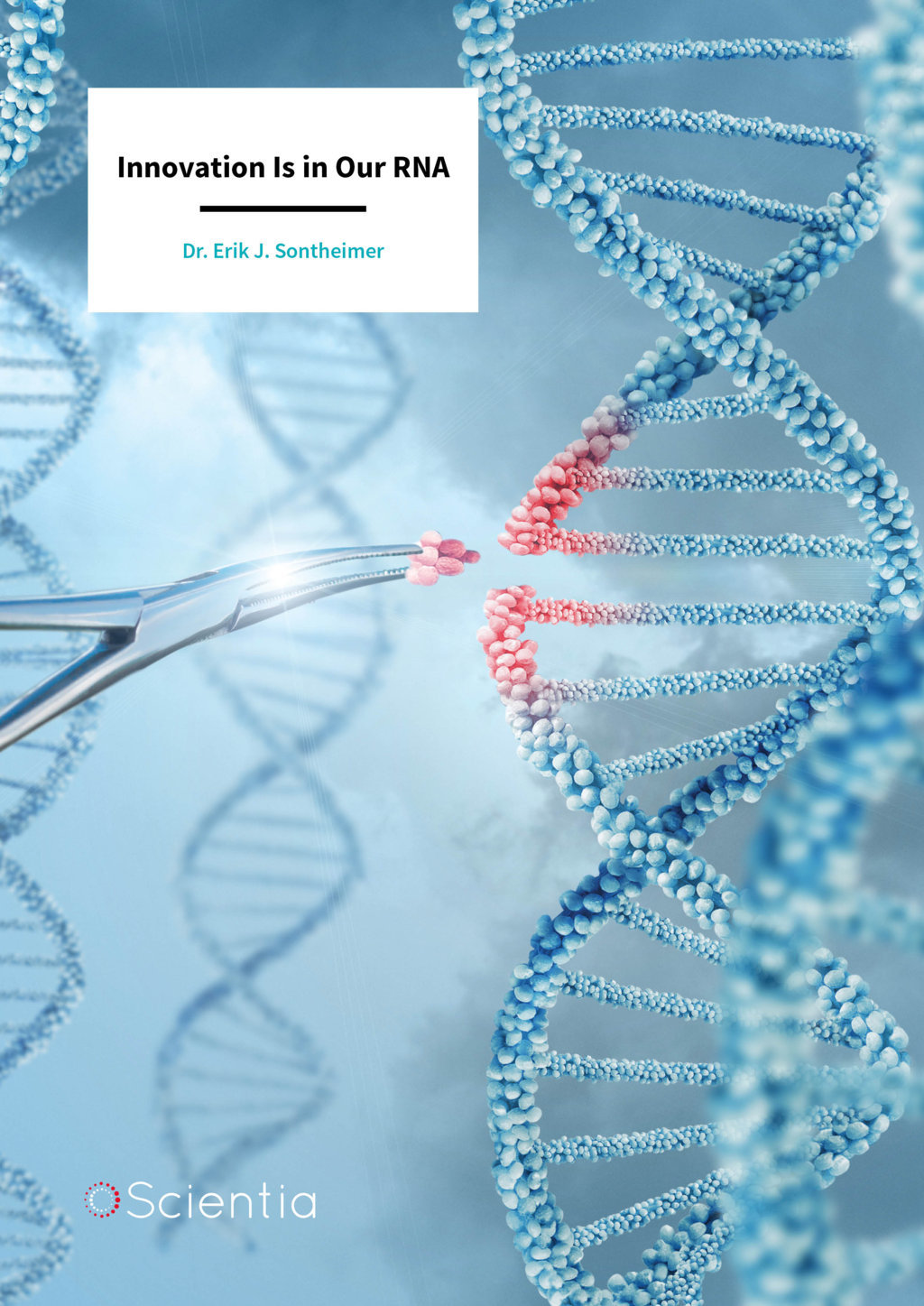
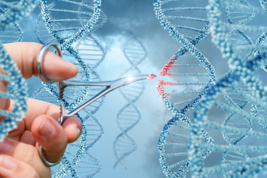
CRISPR-Cas9 systems have become a must-have tool in molecular biology labs the world over for gene editing. Using engineered crRNA guides with Cas9, scientists can make targeted breaks in DNA, which are then fixed by the cell’s natural DNA repair mechanisms. The possibilities are almost endless – insertions of new genes, deletion of unwanted genes, gene replacement and gene activation or inhibition. While conventional genetic engineering has been around for decades, the relative simplicity, ease, and ultra-precision of the DNA scissors that is CRISPR, has made it uniquely powerful for editing genomes within living cells. From being an obscure, niche field of research, CRISPR has revolutionized biology – and the Sontheimer lab has a pioneering place in this revolution.
Staphylococcus is a type of bacteria that may be pathogenic and pose an infection risk to immunocompromised patients in hospitals, and worryingly, it can attain resistance to antibiotics by acquiring foreign DNA. In 2008, Dr. Sontheimer and a postdoctoral fellow in his lab, Luciano Marraffini, investigated CRISPR in Staphylococcus epidermis. They discovered that Staphyloccocus uses a CRISPR sequence to prevent the uptake of foreign DNA – presenting a potential opportunity to fight antibiotic resistance. They were the first to demonstrate that CRISPR interference can directly target DNA, rather than RNA – a highly significant finding because organisms’ genomes are composed of DNA. This discovery paved the way for a wave of genome-editing CRISPR applications. As their paper states: “from a practical standpoint, the ability to direct the specific addressable destruction of DNA…could have considerable functional utility, especially if the system can function outside of its native bacterial context.”
“These advances first pointed the field down the developmental path of genome editing, setting the stage for the recent revolution in RNA-guided genome manipulation,” states Dr. Sontheimer. In January 2009, Dr. Sontheimer submitted a grant application to the NIH proposing to develop CRISPR as an RNA-directed genome editing platform, but the NIH was apparently not ready to recognize the transformative potential of this technology, and the application was not funded. Only a few years later in 2012, this idea proved to be revolutionary.
Dr. Sontheimer’s vision of CRISPR for eukaryotic gene editing is now a reality. Now routinely conducted around the world, the technology allows scientists to manipulate the phenotypes of plants, animals and humans in a relatively straightforward fashion, with almost endless possibilities for life sciences research. To edit the genomes of eukaryotic organisms, the Cas9 protein from the bacterium Streptococcus pyogenes is normally employed. While this is the current standard, it may not necessarily be the best for all applications – and Dr. Sontheimer’s group are testing alternative bacterial Cas proteins for gene editing.
Recently, using the meningococcus bacteria, the research group investigated the application of a new subtype of CRISPR system – the Type II-C CRISPR-Cas9 pathway – which can offer additional capabilities compared to conventional systems. A promising application is therapeutic gene editing in humans, which involves delivering crRNA-Cas complexes to target tissues in viruses, to delete and/or replace defective genes as a cure for genetic diseases. Unlike Cas9 found in Streptococcus pyogenes, most Type II-C Cas9s are sufficiently small that they can be delivered to target tissues by viruses – making them potential candidates for gene therapy. Another challenge for eukaryotic CRISPR applications is that the crRNA-Cas complex may cut and mutate other DNA sequences, and these unintended genome alterations, known as off-target effects can have devastating impacts. Dr. Sontheimer’s team has recently demonstrated that NmeCas9 – the Cas9 protein from the bacterium Neisseria meningitidis – shows unusually high accuracy during human genome editing, without the extensive engineering required for decreasing these off-target effects of Streptococcus pyogenes Cas9, improving its candidacy for use as a targeted therapeutic.
CRISPR is a powerful tool for in vivo gene editing, but must be harnessed and controlled to prevent unwanted interference. For example, off-target effects can be exacerbated by excessive or prolonged Cas9 activity – presenting a safety challenge for therapeutic applications. One possible solution is an off-switch for CRISPR after the desired editing has taken place. Using the NmeCas9 system, Dr. Sontheimer’s group and their collaborators have been the first to discover viral proteins that can stop the activity of Cas9. These so-called anti-CRISPRs are used as Cas9 off-switches and greatly increase our control over genome editing. The Sontheimer lab is investigating the potential of the NmeCas9 system in human therapeutic gene editing, with an emphasis on immunodeficiency, as a means for curing diseases at the genetic level.
Summary
The RNA world is proving to be more diverse than anyone could have possibly imagined a few decades ago. Beyond protein production, RNA has a number of previously unimaginable roles – genetic regulation, signaling, catalysis and even genomic immunity. RNA-based interference can be exploited for cutting-edge biotechnological applications – and nowhere is this more apparent than the recent explosion of CRISPR gene-editing innovation. Dr. Sontheimer’s lab is at the forefront of CRISPR research, and has been the earliest and one of the most vocal advocates of this highly-versatile molecular biology toolkit. Going beyond simply optimizing CRISPR systems for greater control, the lab aims to apply this toolkit for correcting disease-causing genes – therapeutic genomic editing to cure inherited diseases.
Meet the researcher
Dr. Erik J. Sontheimer
RNA Therapeutics Institute
University of Massachusetts Medical School
Worcester, MA
USA
Dr. Erik J. Sontheimer is a Professor in the RNA Therapeutics Institute, and the Program for Molecular Medicine at UMass Medical School. Throughout his academic career, he has pursued his research interests in RNA molecular biology, the roles of RNA in gene expression and regulation, and gene editing. Dr. Sontheimer was awarded his PhD at Yale in 1992, under the tutelage of Dr. Joan Steitz, before gaining a Jane Coffin Childs Postdoctoral Fellowship at the University of Chicago in Dr. Joseph Piccirilli’s laboratory. Between 1999 and 2014, he was on the faculty of the Department of Molecular Biosciences at Northwestern University in Evanston, Illinois, where he rose to the rank of Professor in 2012. In 2007, he began investigating CRISPR-Cas interference mechanisms in pathogenic bacteria, and has since pioneered the optimization of CRISPR-Cas systems for genome editing. In 2014, Dr. Sontheimer moved to the RNA Therapeutics Institute, where his CRISPR research is more geared towards optimization of CRISPR systems for application to therapeutic gene editing.
CONTACT
E: erik.sontheimer@umassmed.edu
T: (+1) 774 455 3639
W: http://www.umassmed.edu/sontheimerlab/home/
REFERENCES
A Pawluk, N Amrani, Y Zhang, B Garcia, Y Hidalgo-Reyes, J Lee, A Edraki, M Shah, EJ Sontheimer, KL Maxwell and AR Davidson, Naturally Occurring Off-Switches for CRISPR-Cas9, Cell, 2016, 167, 1829–1838.
Y Zhang, N Heidrich, BJ Ampattu, CW Gunderson, HS Seifert, C Schoen, J Vogel and EJ Sontheimer, Processing-Independent CRISPR RNAs Limit Natural Transformation in Neisseria Meningitidis, Molecular Cell, 2013, 50, 488–503.
LA Marraffini and EJ Sontheimer, CRISPR Interference Limits Horizontal Gene Transfer in Staphylococci by Targeting DNA, Science, 2008, 322, 1843–45.
JW Pham, JL Pellino, YS Lee, RW Carthew and EJ Sontheimer, A Dicer-2-Dependent 80s Complex Cleaves Targeted mRNAs during RNAi in Drosophila, Cell, 2004, 117, 83–94.
EJ Sontheimer, S Sun and JA Piccirilli, Metal Ion Catalysis during Splicing of Premessenger RNA, Nature, 1997, 388, 801–805.
EJ Sontheimer and JA Steitz, The U5 and U6 Small Nuclear RNAs as Active Site Components of the Spliceosome, Science, 1993, 262, 1989–96.
![]()