Dr Kayvan Najarian – A Revolutionary Approach to Emergency Medical Care
The multiple injuries sustained in a traumatic accident put victims’ lives at risk and can be difficult to diagnose accurately in fast paced emergency room settings. Dr Kayvan Najarian and his team of researchers at the University of Michigan are designing advanced automated trauma decision support systems that can help emergency physicians make critical time-sensitive decisions in life or death situations.
The Trouble with Trauma
Trauma, the medical term that includes the vast range of injuries that might be sustained during an accident such as a car crash, is the leading cause of death for United States citizens under the age of 41. In cases of severe trauma, it is common for a patient to have sustained many injuries in multiple parts of the body, and the lives of these patients often depends on a quick and accurate diagnosis, followed by a treatment plan that prioritizes the most dangerous injuries.
Patients arriving in the emergency room with severe injuries present some of the greatest challenges for medical staff and are at the greatest risk for further complications if a poor diagnosis is made or treatment is delayed. Thus, tools that aid in making these critical decisions can be the difference between life and death.
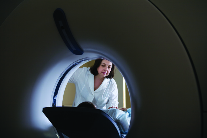
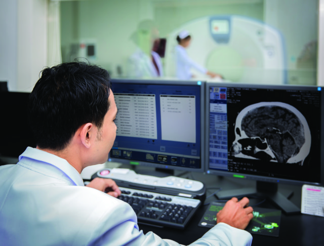
Often doctors are in a rush to obtain as much diagnostic data as possible as quickly as possible, and a Computed Tomography scan or CT scan is often the tool of choice to quickly assess the extent of damage to a patient’s bones and internal organs following an accident. ‘In the majority of traumatic injuries, patients suffer from polytrauma, that is, they often suffer from trauma to multiple systems and parts of the body,’ says Dr Kayvan Najarian, Associate Professor of Emergency Medicine at the University of Michigan. ‘In particular, pelvic and abdominal injuries often accompany brain injuries. Consequently, information contained in head, abdominal, and pelvic CT scans are among the most important medical data available for clinical decision-making.’
During a CT scan, the patient lays inside a special machine that uses x-ray technology to create multiple digital images of ‘slices’ of a patient’s body. When these images are stacked together it creates a 3D model of the inside of the body, allowing doctors to assess internal injuries without making any cuts or employing any invasive techniques.
‘We have developed a computer-assisted decision support system capable of rapidly analyzing large volumes of patient information to generate accurate treatment recommendations and outcome predictions.’
Dr Najarian’s background is in electrical and biomedical engineering, which he began applying to CT images in 2008, when his team worked to develop an improved method of analyzing CT brain scans following a traumatic head injury. Typically, following a CT scan, the multitude of image slices must be carefully aligned before a 3D analysis of the structure can be performed.To speed the analysis of brain CT scans, Dr Najarian’s team created an algorithm to align the image slices more quickly and detect abnormalities. In addition to a clearly defined midline, the brain contains four major fluid-filled cavities called ventricles, where spinal fluid is produced. The algorithm starts by locating the midline, then aligning the location of the ventricles.
E-book

Audio-book

Reference
https://doi.org/10.26320/SCIENTIA123
Share
By comparing to a database of healthy and injured brain scans, the program is then able to suggest potential injury sites to doctors to aid in diagnosis. Dr Najarian and his team are now focused on developing computerized methods of aiding in CT assessments.
‘The main research focus of the Biomedical and Clinical Informatics lab has been the creation of computer-aided decision systems for complex clinical problems, in particular designing automated decision support systems,’ Dr Najarian explains. ‘In collaboration with numerous researchers in engineering, basic sciences, life sciences, as well as clinicians, we identify clinical problems that can be solved by mine and my lab members’ research gains in signal processing, machine learning, and image processing. These systems are designed with the express purpose of translating this research to operational clinical settings.’
Revolutionizing Pelvic Injury Diagnosis
After their success in augmenting CT processing for brain scans, Dr Najarian and his colleagues began to explore other ways that advanced image analysis algorithms could be used to aid in medical decisions and help in emergency trauma situations. Their next point of focus was the pelvis. Due to the proximity of many major organs to the pelvis, pelvic injuries can be some of the most threatening to a patient’s immediate health and are highly associated with increased mortality rates.
Unfortunately, when a patient has sustained many traumatic injuries it is more likely that one will be missed, particularly when the injury is to an internal structure – misdiagnosis of the most critical injury occurs in 16.2% of multiple trauma situations, which increases to 19.6% when there are multiple abdominal and pelvic injuries, and 39.5% when both the pelvis and limbs are involved.
CT scans of the abdomen and pelvis are among the most important tools for assessing the severity of internal injuries but can contain massive amounts of information that are difficult to process through visual inspection alone. The pelvis contains multiple complex bone structures that naturally vary in shape and size between individuals, and the abdomen contains multiple organs that exhibit a similar grey intensity on standard CT scans, making it more difficult to detect edges and abnormalities within these regions.
The first goal of optimizing pelvic CT analysis is to accurately map bone location and placement in relation to the other organs in the pelvic region. In 2009, Dr Najarian and his team developed a novel method of creating 3D maps of bone locations from CT images to enhance bone structure visualization. This innovative method of processing preserves accurate shape and size of bones in the 3D model rendered from 2D CT images. The next step in the process was much more complicated – could the team’s system learn to detect fractures?
The pelvis is a particularly difficult area for a computer to detect fractures in – the bones have complex structures, with a great amount of variation between different people, and many bones that are close together may create gaps that a computer could interpret as a fracture where no fracture exists. Even the trained eye of an experienced emergency room doctor can easily miss pelvic fractures on a CT scan – the complexity of this area fools people and computers alike.
In 2012, Dr Najarian and his team debuted a new version of their pelvic CT analysis software that utilizes a novel algorithm to detect fractures in pelvic bones. The system first identifies and maps bone boundaries, then uses known anatomical data to locate fractures automatically. In 2013, the team updated the system with an even more robust image analysis algorithm that combines data from each CT image to accurately identify fractures, even when patients have more unique natural pelvic shapes.

Beyond Bones: Locating Soft Tissue Damage
Dr Najarian and his team realized that one of the greatest challenges for a system that aids trauma injury diagnosis is locating the soft tissue damage that can often be more deadly than a broken bone. Internal hemorrhaging in the abdomen or pelvis is one of the greatest concerns for doctors, as it is the leading cause of death within 24 hours of an accident.
Detecting hemorrhages quickly after trauma is critical to patient survival. Yet historically, it has been highly time consuming for doctors to locate hemorrhaging manually from CT images, delaying the time-sensitive decision-making process that can mean life or death after a traumatic accident. In 2012, the team developed a new method of automated hemorrhage detection and boundary mapping from CT data.
Their system starts by analyzing each slice of a CT scan for unusual blood pooling – if a hemorrhage is found in a given slice, the system then uses it as a starting point to build a 3D model of the hemorrhage boundaries within the pelvis. The software is able to compare this with known anatomical data to identify normal areas of blood flow, such as large veins, and reduce false positives. The use of this technology drastically reduces the amount of time it takes doctors to identify and diagnose internal bleeding, saving crucial time in a trauma patient’s treatment.
Another critical challenge for CT based diagnosis is differentiating and diagnosing damage to the many organs in the pelvic region. Solid organs, such as the spleen and liver, look very similar on the x-ray imaging technology employed by standard CT machines. As with other areas of the pelvis, identifying organs and damage can be difficult for both the human eye and a machine.
In 2015, Dr Najarian and his team demonstrated that their software system could be trained to accurately assess spleen health from a CT scan. Using a unique edge-finding technique, the system is able to differentiate between minute variations in shades of grey to find the boundaries of the spleen and identify lacerations and hemorrhaging in the organ. This technology can be applied to other organs, and when combined with the lab’s other imaging algorithms forms a powerful system to aid emergency physicians.
Dr Najarian describes, ‘we have developed a computer-assisted decision support system capable of rapidly analyzing large volumes of patient information to generate accurate treatment recommendations and outcome predictions. This patented technology intends to provide a near-real-time system to assist medical caregiver decision-making in traumatic pelvic and abdominal injury cases by extracting information from all available patient data, especially CT scans, and integrating the extracted features using machine learning to generate prediction, warning, and treatment recommendations at every stage of patient care.’
Transforming the Use of CT in Trauma Care
Working with emergency medicine researchers at Virginia Commonwealth University and the University of Michigan, along with the Michigan Center for Integrative Research in Critical Care, Dr Najarian and his team are now piloting the use of their automated trauma decision support systems in real life hospital settings. Through the use of multiple advanced algorithms, the system is able to first analyze CT scan data to detect injury, and then combine its findings with patient demographic data to make predictions for the best course of action for that individual patient’s treatment.
The system accurately makes critical predictions such as how many days the patient will need to stay in the intensive care unit, and the likely scenarios for care following their release. Dr Najarian states, ‘the next step for the polytrauma project is to develop a fully functional polytrauma decision support system suitable for commercialization.’ The team hopes to release the system for use worldwide in the near future. With further development, the system could streamline emergency room and intensive care unit functions, such as how much plasma a patient will need, the potential success of various surgical outcomes, along with medications and dosage requirements.
In addition to the trauma decision support system, Dr Najarian’s team is working on other technologies that could revolutionize healthcare, such as an automobile system that could predict the onset of a severe cardiac event and alert a driver prior to complications that could compromise driving safety.
They are also working on a program to automate the interpretation of coronary angiogram videos visualizing heart blood vessels and building an international dataset of such videos for medical research use. The tools developed by Dr Najarian and his team are bringing cutting-edge innovations to patient care and saving lives in the most difficult of situations.
Meet the researcher

Dr Kayvan Najarian
Department of Computational Medicine and Bioinformatics
Department of Emergency Medicine
Michigan Center for Integrative Research in Critical Care
University of Michigan
Ann Arbor, MI
USA
Dr Kayvan Najarian began his education in Iran at the Sharif University of Technology, earning a bachelor’s degree in Electrical & Computer Engineering in 1990. He completed a master’s degree in Biomedical Engineering at the Amirkabir University of Technology in Tehran, Iran in 1994, and then continued on to earn his doctorate in Electrical & Computer Engineering from the University of British Columbia, Vancouver, Canada in 2000. He served as an Assistant Professor of Computer Science at the University of North Carolina from 2001 until 2007, before taking a faculty position at Virginia Commonwealth University in 2007, where he earned tenure in the Computer Science Department and served as the Director of the Biomedical Signal and Image Processing Group. In 2013 he moved his laboratory to the University of Michigan to serve as an Associate Professor in the Department of Emergency Medicine, and the Department of Computational Medicine and Bioinformatics. Here, he is also the Director of Data Science for the Michigan Center for Integrative Research in Critical Care. Dr Najarian has dedicated his career to using the tools of computer science to automate and improve all aspects of human health and medical care.
CONTACT
E: kayvan@umich.edu
T: (+1) 734 763 3924
W: http://najarianlab.ccmb.med.umich.edu/
FUNDING
US National Science Foundation
National Institutes of Health
US Department of Defense
American Heart Association
Toyota Motor Company
REFERENCES
W Chen, R Smith, S-Y Ji, KR Ward and K Najarian, Automated ventricular systems segmentation in brain CT images by combining low-level segmentation and high-level template matching, BMC Medical Informatics and Decision Making, 2009, 9, S4–14.
P Davuluri, J Wu, Y Tang, CH Cockrell, KR Ward, K Najarian, and RH Hargraves, Hemorrhage Detection and Segmentation in Traumatic Pelvic Injuries, Computational and Mathematical Methods in Medicine, 2012, 6, 1–12.
SM Reza Soroushmehr, P Davuluri, S Molaei, RH Hargraves, Y Tang, CH Cockrell, et al. Spleen Segmentation and Assessment in CT Images for Traumatic Abdominal Injuries, Journal of Medical Systems, 2015, 39, 87.
S Vasilache, K Ward, C Cockrell, J Ha, and K Najarian, K, Unified wavelet and Gaussian filtering for segmentation of CT images; application in segmentation of bone in pelvic CT images, BMC Medical Informatics and Decision Making, 2009, 9, S8–8.
J Wu, A Belle, RH Hargraves, C Cockrell, Y Tang, Y and K Najarian, Bone segmentation and 3D visualization of CT images for traumatic pelvic injuries, International Journal of Imaging Systems and Technology, 2014, 24, 29–38.
J Wu, P Davuluri, KR Ward, C Cockrell, R Hobson, and K Najarian, Fracture Detection in Traumatic Pelvic CT Images, International Journal of Biomedical Imaging, 2012, 23, 1–10.