
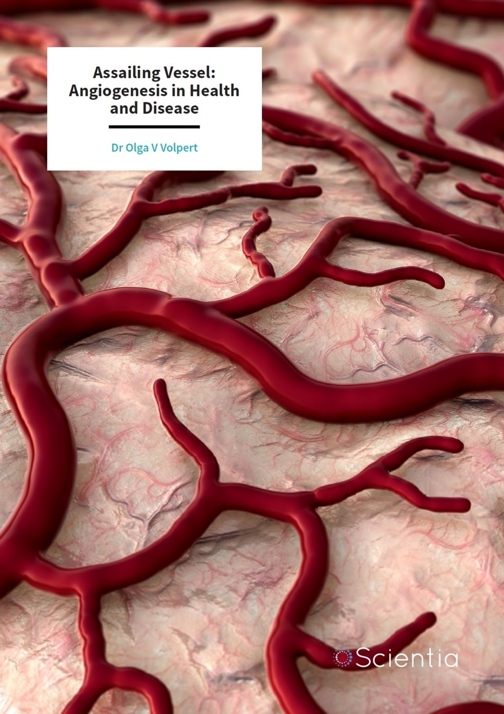
Dr Olga Volpert – Assailing Vessel: Angiogenesis in Health and Disease
Angiogenesis – the formation of new blood vessels – is a major component of the progression of several diseases – especially cancer. Dr Olga Volpert at the University of Texas MD Anderson Cancer Center works on elucidating the molecular mechanisms of abnormal angiogenesis, for the development of novel therapies.
Targeting Angiogenesis in Cancer
The cardiovascular system – this network of veins, capillaries, and arteries, powered by the heart and supported by tissue fluid that bathes cells – provides cells with vital nutrients and oxygen, while removing waste products. In the embryo, primitive blood lacunae give rise to the first blood vessels. Hematopoietic cells then give rise to the primitive endothelial (vascular) cells that proliferate and coalesce to form the initial vascular network in a process termed vasculogenesis. This vascular network then expands by sprouting new capillaries (angiogenesis) in response to pro-angiogenic factors. In healthy adults, angiogenesis is rare and the vascular network of veins, capillaries and arteries is fairly stable – thanks in part to a range of anti-angiogenesis factors in cells and blood that counteract angiogenic stimuli. However, this delicate balance between angiogenic inhibitors and stimuli can be dysregulated, with associated changes in the vasculature and blood supply to the tissue. Dysregulated angiogenesis is implicated in a number of diseases.
In particular, angiogenesis is a major component of cancer. Cell division is normally a tightly regulated and orchestrated process, but in cancer, this proliferation escalates due to genetic events known as oncogenesis, and abnormal cells divide rapidly and uncontrollably to form tumours. Cancerous cells, just like normal cells, depend on a blood supply to deliver nutrients and oxygen, and remove waste products – perhaps more so than normal cells due to their abnormally high growth and metabolic activity. Once tumours grow beyond a few millimetres in diameter, they no longer rely on diffusion and promote the formation of their own network of capillaries, allowing them to overcome the growth constraints. Angiogenesis is often a pre-requisite to cancer metastasis – an advanced stage of cancer progression in which malignant cells migrate to other parts of the body – spreading the cancer. Thus, early diagnosis is important to improve patient survival outcomes. In contrast, ischemia refers to a range of conditions characterised by insufficient blood supply – causing organs to be starved of oxygen – which can lead to irreversible tissue damage and necrosis. Thus, therapies that stimulate angiogenesis have been advocated as a means mitigate against ischemic damage and improve blood supply.
E-book
Reference
https://doi.org/10.26320/SCIENTIA69
Share
Due to the importance of angiogenesis in cancer, suppression of angiogenesis is a target of cancer treatment. Vascular epithelial growth factor (VEGF) is a major activator of angiogenesis, and the VEGF pathway is highly active in a number of cancers. Hence, the majority of anti-angiogenic cancer therapies block the VEGF pathway. Bevacizumab, an anti-VEGF monoclonal antibody, approved by the FDA in 2004, has become a standard component of cancer therapy – alone or in combination with chemotherapy – and was soon followed by a wave of other VEGF inhibitor drugs. Notwithstanding, VEGF-targeting anti-cancer drugs typically elicit modest clinical improvements and may have adverse side effects including hypertension, thromboembolism, renal failure and intestinal perforations – calling for the development of more effective and safer anti-angiogenic therapies.
Dr Olga Volpert, Associate Professor at the University of Texas MD Anderson Cancer Center, works on elucidating the mechanisms of pathological angiogenesis, especially cancer angiogenesis – and applying these to developing novel therapeutic approaches that target the cardiovascular system.

Controlling Angiogenesis with MicroRNAs
New microRNA-targeting therapeutics may be a safer, more effective alternative to current VEGF inhibitors – as they provide a far subtler degree of regulation at the post-transcriptional level. MicroRNAs (miRNAs) are small non-coding RNA molecules 18–25 nucleotides in length, involved in regulating gene expression. MiRNA binds to transcribed mRNA, negatively regulating its expression, through mediating mRNA cleavage or translation repression. It is recognised that miRNAs are involved in regulating several cellular functions, including cell migration, apoptosis, signal transduction, metabolic pathways – and dysfunctions, such as tumorigenesis, tumour growth and angiogenesis. Expression of certain miRNAs are associated with the progression or suppression of cancer.
MiR-27b is a dysregulated miRNA that can act either promote or suppress cancer progression. In many cases, miR-27b stimulates angiogenesis. Previous studies carried out by Dr Volpert and colleagues have demonstrated that miR-27b promotes capillary sprouting and influences the fate of endothelial tip cells (which direct the growth of new capillary sprouts and are critical for angiogenesis) by suppressing the production of the anti-angiogenic factors Delta-like ligand 4 (DII4) and Sprouty-2 (Spry2) – essential for vascular stasis and early segregation of veins and arteries.
Recent studies by Dr Volpert and her colleagues, using Lewis lung carcinoma (LLC1), mouse macrophage cell lines and mouse models have indicated the potential of targeting miR-27b as a therapeutic intervention for cancer and other diseases involving dysregulated angiogenesis. Using a synthetic oligonucleotide, antagomir, that specifically binds and destroys miR-27b – called anti-miR-27b – they demonstrated that inhibiting miR-27b suppresses cancer growth and angiogenesis in mice. Conversely, they used a synthetic miR-27b mimic to investigate miR-27b as a therapy for limb ischemia (a model of diabetic ulcers) and myocardial infarction (heart attack) – both vascular disorders. They demonstrated that the miR-27b mimic increases angiogenesis and tissue repair, and minimises tissue damage in ischemic limb and in the heart wall.
These findings demonstrate that targeting miR-27b can be used as a therapeutic strategy in cancer, and restoring miR-27b is beneficial in vascular disorders. Current interventions for treating ischemic diseases involve inserting mechanical stents to increase blood flow through diseased vessels. While stem cell and gene therapies have been advocated to treat ischemic disease, they have thus far not entered the clinic. MiRNA approaches such as delivering miR-27b to diseased tissue may be a safer, more effective alternative. Further preclinical and clinical studies need to be conducted to address this – in particular, successful delivery of active oligonucleotides to tissues remains a major bottleneck.

An Angiogenic Inhibitor from an Unlikely Source
Sunlight-induced skin damage has been in the public health spotlight since the 1980s ozone layer depletion scare. Long-term exposure to ultraviolet B (UVB) radiation in sunlight is a risk factor for both non-melanoma and melanoma skin cancers. The incidence of these skin cancers is increasing, with 2–3 million non-melanoma skin cancers and 132,000 melanoma cases occurring globally each year. Skin can show the signs of photodamage even after short exposures to UVB irradiation. As with other cancers, angiogenesis plays major role in skin cancer progression, and UVB-induced vascular changes in the skin have been attributed to increased activity of pro-angiogenic cytokines and chemokines, including VEGF, Cox-2, basic fibroblast growth factor (bFGF), and interleukin-8 (IL-8).
Thrombospondin-1 (TSP1), a large glycoprotein abundant in healthy adult tissues is a major inhibitor of angiogenesis. Loss of TSP1 has been implicated in the progression of many cancers including breast and colon carcinomas and skin cancers. In the skin, TSP1 is expressed by epidermal keratinocytes, and acts as a natural gatekeeper of the vascular changes associated with progression of cancer. Therefore, TSP-1 seems an obvious candidate for cancer prevention or treatment. It can be either administered TSP-1 as a biologic therapeutic; however, it is a very large protein with complex functions and difficult to produce for clinical purposes. What if there was a compound that impedes subcutaneous angiogenesis by elevating expression of TSP-1 in skin, potentially with anti-tumorigenic effects? There may be such a compound – and it’s from an unlikely source.
Chamomile flowers have been revered since antiquity, for their purported healing properties. A compound naturally found in chamomile, apigenin, has been found to have potent chemoprotective properties against UVB-induced skin cancer. As well as cytotoxic and cytostatic effects (potentially useful for arresting the growth of rapidly proliferating tumour cells), apigenin inhibits angiogenesis. Peptides that mimic anti-angiogenic effects of TSP1, such as ABT-898 have shown similar anti-angiogenic and anti-tumorigenic properties.
Using cultures of human and mouse keratinocyte cell lines and mouse models, Dr Volpert and her colleagues carried out studies confirming the anti-angiogenic effects of apigenin, and demonstrated that apigenin mitigates TSP1 loss in the UVB-exposed skin – the first known report of apigenin being directly involved in the regulation of endogenous anti-angiogenic factors. They found that TSP1 levels dramatically decreased with UVB exposure, although with apigenin, before or after UVB irradiation, restored normal TSP1 levels. They also found that like apigenin, ABT-898 inhibited the activity of Cox-2 and VEGF and reduced UVB-induced vascular effects associated with onset of skin cancer – angiogenesis and epidermal thickening. These studies strongly suggest the potent therapeutic benefits of apigenin and its mimetics – as a potential therapy or preventative measure for skin cancer, and provide insights into their protective mechanisms.
Non-Malignant Fibroblasts in Melanoma
Malignant tumours are composed of neoplastic (rapidly dividing) cells. There are also non-malignant cells in the tumour microenvironment including fibroblasts, endothelial cells, immune cells and bone marrow-derived cells. These are not innocent bystanders – they are just as critical to cancer progression. It is now recognised that there is ‘crosstalk’ between malignant and non-malignant cells – causing these non-malignant cells to switch to tumour-associated phenotypes.
Fibroblasts are important cells in skin physiology. However, cancer-associated fibroblasts are a major constituent of the tumour microenvironment in melanoma (skin cancer) – and are thought to be involved in making tumours resistant to chemotherapy drugs. The emergence of the tumour-associated phenotype is driven by environmental stresses and a host of cytokines. Cancer-associated fibroblasts support tumour progression by releasing a range of growth factors – basic fibroblast growth factor (bFGF), epithelial growth factor and hepatocyte growth factor (HGF) – and angiogenic factors – VEGF, interleukin-8 and stromal-derived factor 1 (SDF-1). In healthy connective tissue, fibroblasts synthesise collagen and the extracellular matrix important for providing a structural scaffold – but in cancer, this scaffold supports tumour progression.
Pigment epithelial-derived factor (PEDF) is a secreted glycoprotein with anti-angiogenic and tumour-suppressing properties. Dr Volpert’s team previously discovered that loss of PEDF expression in melanocytes enables melanomas to acquire an invasive metastatic phenotype. However, the role of PEDF expression in fibroblasts in suppressing skin cancer, and the impact of the extracellular matrix, are not well known. To investigate this, Dr Volpert and her colleagues recently carried out studies, which included PEDF knock-out human dermal fibroblasts and mice. Accordingly, PEDF-depleted fibroblasts were found to promote tumour growth and PEDF-knockout mice were more susceptible to melanoma metastasis. More interestingly – cells champion their own proliferation by suppressing PEDF production in fibroblasts, which also appears to expedite the transition of healthy fibroblasts to a cancer-associated state. These studies suggest that the promotion of PEDF expression by fibroblasts – or external delivery of PEDF and peptides based on PEDF as targeted therapies – can be an affective therapeutic approach to treating melanoma.
Senescence as a Potential Therapeutic Approach for Cancer
Cellular senescence is a state in which cells cease to divide – though still remain metabolically active. A very obvious – yet largely uninvestigated chemotherapy approach – could be inducing senescence in cancer cells. Dr Volpert and her colleagues are investigating this cellular phenomenon in prostate cancer. Prostate tumours overexpress androgen receptors, and previous studies suggest that activating this receptor stimulates tumour proliferation and inhibits apoptosis – though this is far from conclusive. Dr Volpert and colleagues have recently carried out studies in which prostate cancer cells undergo cellular senescence through continuous activation of androgen receptors. These pioneering studies suggest the potential of using androgen receptor-stimulating compounds in chemotherapy to stop the growth of tumours by inducing senescence. However, much research needs to be done first before clinical studies – particularly to resolve the previous apparent contradictory findings in the pro- or anti-proliferative effects of AR stimulation.
Angiogenesis Inhibitors Packaged in Exosomes
Exosomes are tiny physiological vesicles (50–150 nanometres in diameter) formed through endosomal pathway and released by all cells in the body and travel in biological fluids, like serum and urine. Studies in the past decade have identified exosomes as the vehicles in critical cell-to-cell and cross-tissue communications. These natural nanovesicles carry proteins, nucleic acids and lipids and alter the biology of the cells they enter. Most studies show that exosomes released by cancer cells promote metastasis by creating permissive environments at the sites of their arrival (metastatic niches).
In a recent study, Dr Volpert and colleagues found that at an early stage, melanoma cells alert the immune system of the host to the presence of metastasis by sending out exosomes that activate cells called patrolling monocytes, which then seek out cancer cells in the blood vessels at a distant site. Working with melanoma as a model, Dr Volpert showed that one of such exosome-associated activators of patrolling monocytes in pigment epithelium-derived factor (PEDF) – a known angiogenesis inhibitor – which in this case ‘doubles’ as an activator of immune response.
Summary
The FDA approval of bevacizumab in 2004 was a milestone in cancer treatment – bringing the importance of dysregulated angiogenesis to the spotlight. Thirteen years later, there is much about angiogenesis that we do not fully understand – and Dr Volpert and her team are trying to elucidate the complex molecular agents and mechanisms involved. They hope that the insights from this pioneering research will lead to novel approaches to the treatment of cancer and other diseases such as macular degeneration.
Meet the researcher

Dr Olga V Volpert
MD Anderson Cancer Center
University of Texas
Houston, TX
USA
Dr Olga Volpert graduated with a Master’s degree in General Microbiology at Lomonosov Moscow State University (MSU) in 1983. In 2000, she began her tenure as Associate Professor at Northwestern University, where she carried out research for nearly two decades. She has recently moved her lab to the MD Anderson Cancer Center, University of Texas. Dr Volpert’s research themes include the molecular processes underpinning angiogenesis (blood vessel formation) in cancer and eye diseases, the preclinical development of anti-angiogenic agents, and angiogenic factors as biomarkers of disease progression. Her current research interests include: development of short anti-angiogenic peptide derivatives of Pigment Epithelial-derived Factor (PEDF) and Thrombospondin-1 (TSP1) for sustained release in the treatment of age-related macular degeneration, ovarian cancer and renal failure; exosomes as the negative regulators of tumour angiogenesis, immune suppression and metastasis; and the role of exosomal PEDF as a predictor of melanoma outcome.
CONTACT
E: OVolpert@mdanderson.org
T: (+1) 832 7501521
W: https://mdanderson.influuent.utsystem.edu/en/persons/olga-volpert
REFERENCES
NG Nwani, ML Deguiz, B Jimenez, E Vinokour, O Dubrovskyi, A Ugolkov, AP Mazar and OV Volpert, Melanoma Cells Block PEDF Production in Fibroblasts to Induce the Tumor-Promoting Phenotype of Cancer-Associated Fibroblasts, Cancer Research, 2016, 76, 2265–76.
D Veliceasa, D Biyashev, G Qin, S Misener, AR Mackie, R Kishore and OV Volpert, Therapeutic Manipulation of Angiogenesis with miR-27b, Vascular Cell, 2015, 7, 6.
X Tong, S Mirzoeva, D Veliceasa, BB. Bridgeman, P Fitchev, ML Cornwell, SE Crawford, JC Pelling and OV Volpert, Chemopreventive Apigenin Controls UVB-Induced Cutaneous Proliferation and Angiogenesis through HuR and Thrombospondin-1, Oncotarget, 2014, 5 11413–27.
D Biyashev, D Veliceasa, J Topczewski, JM Topczewska, I Mizgirev, E Vinokour, AL Reddi, JD Licht, SY Revskoy, and OV Volpert, miR-27b Controls Venous Specification and Tip Cell Fate, Blood, 2012, 119, 2679–87.
Y Mirochnik, D Veliceasa, L Williams, K Maxwell, A Yemelyanov, I Budunova and OV Volpert, Androgen Receptor Drives Cellular Senescence, PloS One, 2012, 7, e31052.
JL Orgaz et al., Loss of Pigment Epithelium-Derived Factor Enables Migration, Invasion and Metastatic Spread of Human Melanoma, Oncogene, 2009, 28, 4147–61.
![]()