
Tan A. Ince, MD, PhD – Fighting Cancer by Keeping Cancer Cells Alive
Pathologist and cancer researcher Professor Tan Ince and his colleagues at the University of Miami investigate human cancers by developing cell culture media to grow cancer cells and normal cells to better study the relationship between the two.

How Do You Study a Malignant Invader?
As the U.S. President – played by Bill Pullman – found out unexpectedly in the midst of an alien invasion in the epic Independence Day, specimens of the alien force had been sequestered in the fabled Area 51 somewhere in the Southwest American desert. The aliens were poked and prodded; their capabilities were studied and reverse engineered. Scientists learned a lot from these captured aliens and this knowledge ultimately helped defeat the invaders. Study the invader and find ways to defeat it – discover its weaknesses, strengths and strategy. Perhaps this is all an allegory or an analogy for the life’s work of Professor Tan Ince, cancer researcher and pathologist from University of Miami Miller School of Medicine in Miami.
For almost twenty years, Professor Ince and colleagues have been researching ways to efficiently grow cancer cells for examination and study, in the hopes of finding new and improved therapies for this most terrifying of diseases. Early in his career, Professor Ince developed a new cell culture nutrient medium that is now widely used to grow human breast and ovary cells. As a pathologist he can diagnose cancer cells taken from patients at surgery or at biopsy. As a cancer researcher, he tirelessly searches for ways to keep those cells alive to discover what they can tell him about the cancer growing inside his patient and find ways of defeating it.
Cancer is a Word – Cancers are a Diverse Population
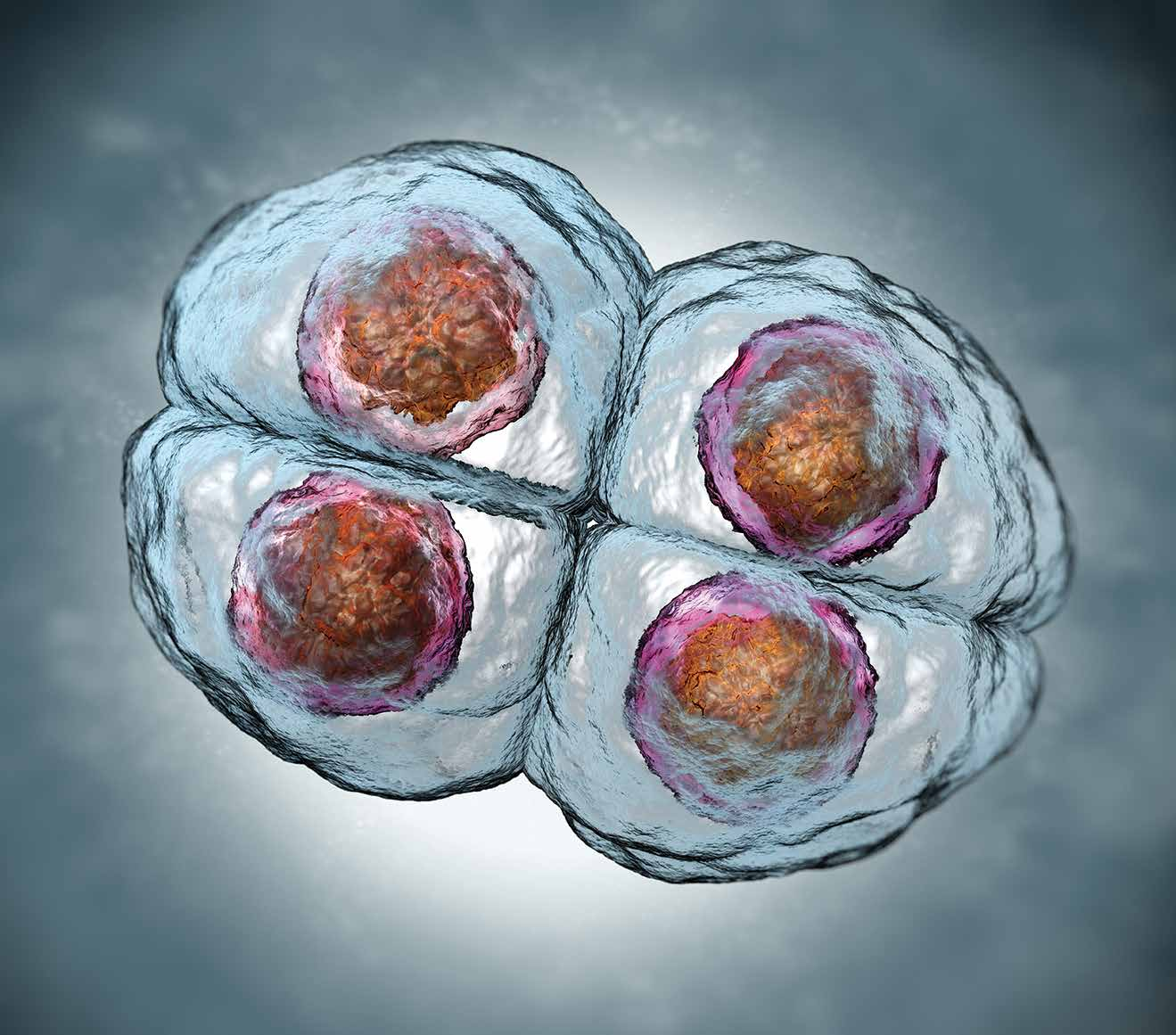
In 1951, a cancer patient, Henrietta Lacks, became immortal. Ms Lacks suffered from cervical cancer and unfortunately succumbed to her disease in October of 1951. However, cells taken from her tumour were successfully cultured, resulting in the socalled HeLa cell line – the first human cell line established in the laboratory – that has since been used extensively for scientific research. The cells from Ms Lacks’ tumour had mutated so much – a fact later discovered to be due to infection with human papillomavirus B-18 – that the cells were immortal and could be used for experiments that were impossible with normal cells. To this day, HeLa cells are used in laboratories around the world. Perhaps ironically, over the decades that the HeLa cell line was used for various and important scientific research, including the testing of Jonas Salk’s first human polio vaccine, the cells have contaminated other cell lines in laboratories worldwide, corrupting research done in laboratory after laboratory. Up to 20% of other cell lines may be contaminated by HeLa cells. Apparently cancers are not only invasive in their original human victims. But that isn’t the only problem with cell lines like HeLa.
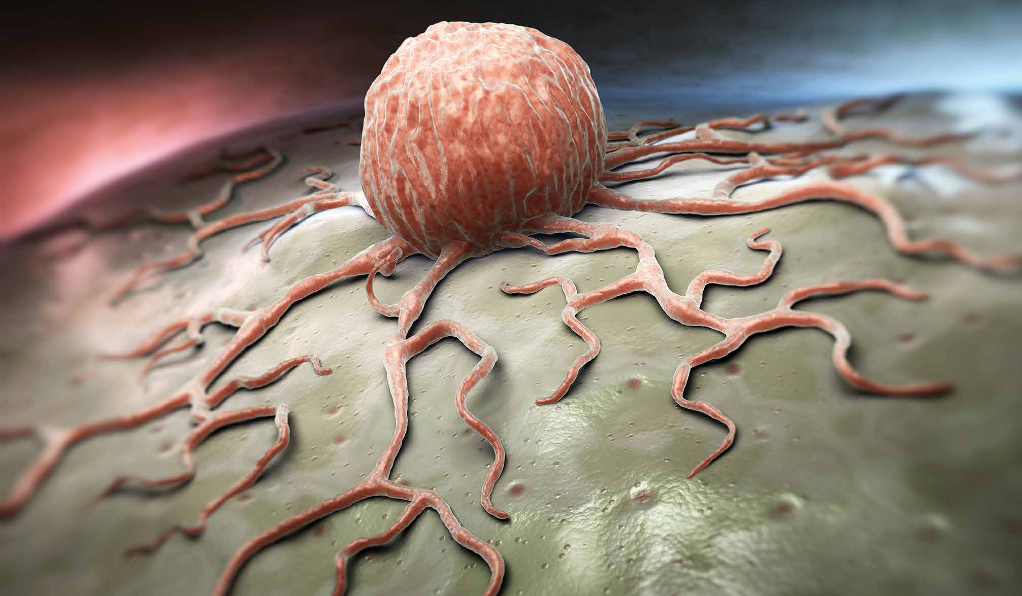
In the decades since the establishment of the HeLa line, human tumour cell lines have had a profound effect on cancer research and led to the development of a variety of human cancer therapies. Carcinomas grow uncontrollably in the body. But the malignant cells are often paradoxically difficult to grow in cell culture, unlike HeLa. A reliable cell line model that predicts patient response to various therapies would be of great value in the testing of new drugs for individualised therapy of tumour patients. But in spite of the many decades of improvements in methods for establishing cancer cell lines, it is quite difficult to routinely establish reliable, permanent cell lines from primary human tumours on a regular basis. This limits the number and diversity of cell lines scientists have at their disposal. As well, with many tumour types only very high-grade specimens have yielded cell lines. Thus, available cell lines do not accurately reflect the true spectrum of tumours encountered clinically. Patients with lower grade tumours are effectively out of luck if their tumour does not readily grow in cell culture.
Another problem is that the origins of many of the available tumour cell lines are unclear. What tumour or even what tissue did they come from? This is the result of a lack of ‘fingerprinting’ technology – such as DNA analysis – that was able to verify identity when the lines were developed. Also, the original tumour usually isn’t available for analysis with modern DNA sequencing procedures. What scientists need is a more efficient method of establishing fresh human tumours in culture to indicate and reflect the heterogeneity of the human tumours from which they are derived. If you are treating a patient with a certain type of breast cancer, you want to experiment on a cell culture from that type of cancer. This would give a more logical experimental model for drug testing, for example.
In fact, Professor Ince and fellow researchers wrote in Nature Communications about their development of a cell culture medium that allowed them to routinely establish cell lines from different subtypes of human ovarian cancers with over 95% efficiency. They described 25 new ovarian tumour cell lines that retained the genetics, histopathology and molecular features of the patient’s original tumours. Importantly for therapeutic strategies, the molecular profile and drug response of these tumour cell lines correlated with distinct groups of primary tumours with different outcomes. In other words, patient outcomes were related to the different tumour types Ince could grow in cell culture. Using a tumour cell line that is not the same type – or even the same grade or malignancy – as the patient’s tumour doesn’t give the correct result.
These tumour cell lines that Professor Ince derived represent a significantly improved experimental system to study human tumour pathophysiology and ultimately response to therapy. Using ‘standard’ cell culture, such as the historic HeLa line, just doesn’t apply to all tumours. Patients need more specificity for a proper chance at some good result. Professor Ince and his team keep working toward that end in a variety of ways.
For almost twenty years, Professor Ince and his colleagues have been researching ways to efficiently grow cancer cells so they can be examined and studied, in the hopes of finding new and improved therapies for this most terrifying of diseases.
Relating the Normal to the Abnormal
To understand the pathophysiology of a disease and most appropriately determine treatment choices for a given cancer, the patient’s doctor needs accurate classification of that cancer. For blood cell malignancies such as leukaemia, a classification scheme based on the phenotypic similarity between cancer cells and normal cells has been successfully used to define subtypes of the cancer. There is lymphocytic leukaemia, derived from lymphocyte cells, granulocytic leukaemia, derived from the other white cells, and so on. Beyond that, the use of normal cell types as a reference by which to classify solid tumours – such as carcinomas or sarcomas – has not been widely practiced, due, in part, to a more limited understanding of solid tumour cell differentiation. Professor Ince and his colleagues have tried to bridge this gap in understanding and basically develop a new paradigm in understanding of solid tumours. They looked at breast cancer – a very common cancer – and tried to get a number of breast cancer tumour types that make for a more diverse possibility for research into therapy of those types of tumours.
Breast cancer is usually a cancer of epithelial cells, the cell type that covers most of the internal and external surfaces of the body and its organs. To get a better handle on the subtypes of epithelial cells comprising the breast epithelium, Professor Ince performed a systematic analysis of a large group of breast epithelial markers in more than 15,000 normal breast cells. This exhaustive study identified eleven differentiation states for the normal breast cells. He used this information to classify breast tumours based on normal cell types into four major subtypes that were differentiated from each other by their metabolism of vitamin D, androgen (male hormone), and oestrogen (female hormone) receptor expression. In other words, some cells had receptors that bonded to vitamin D and some did not. Some had receptors that bonded to oestrogen or androgen and some did not. In this way he distinguished cells based on their receptor status for these three substances.
Professor Ince then looked at cells from 3,157 human breast tumours and found that these hormone receptor subtypes were distinct from the current classification scheme, which is based on oestrogen receptor, progesterone receptor, and human epidermal growth factor receptor 2. Importantly, he found that patient outcomes were better when tumours expressed all three hormone receptors (which he called subtype HR3) and worst when they expressed none of the receptors (called subtype HR0). Putting the whole picture together, these data provide a practical classification scheme associated with actual differences in patient survival and provided doctors insights into possible treatment of these breast tumours. Basically, the more specific you can be regarding the identity of the cancer, the better the chance at treating it. Know your enemy is apparently good advice.
Looking at What’s Real
Professor Ince’s research started in the universe of what was already known, like the relatively unique HeLa line. But he has continued to search for what’s real – tumours related to the real tissue from which they arose. It’s not about a distant and unrelated tumour line from a patient who died half a century ago. It’s about examining cells of the same type as the cells your patient is affected with. To that end, Professor Ince recently suggested a new approach to classification of breast cancer, based on his vast experience with specific cell lines developed from specific tumours and related to specific parent cell types.
While current classification systems for breast cancer are based on expression of prognostic and predictive biomarkers, Professor Ince proposes a hypothesis-based ontological breast cancer classification modelled upon the taxonomy of species as the evolutionary biologist sees it. His approach takes the normal breast epithelial cell types and differentiation lineages as the gold standard to classify tumours arising from breast tissue. In other words, relate the malignant cell type with the normal cell type that presumably parented it.
Professor Ince took his prior research – demonstrating at least eleven previously undefined normal cell types in human breast epithelium and that each breast carcinoma is related to one of these normal cell types – and found that triple negative breast cancers do not have a ‘basallike’ phenotype. The breast cancer cells that were negative for receptors for vitamin D, oestrogen and androgen did not have a ‘normal’ parent cell. Cells from normal tissue never lack all of these receptors. The triple negative tumours must be pretty unique. Normal breast epithelial cells exist in four novel hormonal differentiation states and almost all human breast tumours fall under one of these hormonal differentiation states. This can have significant survival differences, since you can tailor therapy to include, say, oestrogen receptor drugs or androgen receptor drugs. This real-life classification scheme can provide rational treatment guidance and an alternative approach for understanding tumour physiology and tumour classification. And Professor Ince means to follow his classification theory even to the deep molecular level.
Hot off the presses in the journal Oncogene, for example, Professor Ince’s team reported a study of histone deacetylases (HDAC) in breast and ovarian cancer specimens. HDAC are enzymes that help regulate DNA expression by modifying histones, molecules that wrap around and control DNA activity. What he found was that two specific types, HDAC1 and HDAC7, are vital to the function of cancer stem cells, those cancer cells that allow tumours to grow and spread. This means that already existing drugs that inhibit HDAC can rationally be tried on these tumours. Again, classifying tumours by HDAC content can lead to more specific therapeutic choices. In this work Professor Ince has revealed another bit of information on the alien invaders to help in our fight against them!
The epitome of Professor Ince’s research would be his proposal of a stepwise classification system that puts tumours into diagnostic categories based on their distinct tissue of origin, cell-of-origin and differentiation lineage – what he calls ‘lineage based classifications’. After defining uniform lineage based classes, he proposes to use molecular and genetic classifiers – like the presence of HDAC1 and HDAC7 – to distinguish prognostic subsets within each lineage. In other words, in the future he plans to narrow the classifications down so much that if you have this or that cancer, there will be a classification relative to your tumour – we could have a therapy for you.
Meet the researcher

Tan A. Ince, MD, PhD
Associate Professor of Pathology
University of Miami Miller School of Medicine
Miami, Florida, USA
Professor Tan Ince received his MD from Hacettepe University School of Medicine in Ankara, Turkey, in 1988. In 1996 he received his PhD in Molecular Pharmacology from Cornell University in New York City. He then completed a residency in Anatomic Pathology at Massachusetts General Hospital and Harvard Medical School, followed by a fellowship in Women’s and Perinatal Pathology at the Brigham and Women’s Hospital there. Professor Ince was a visiting clinical scientist at the Massachusetts Institute of Technology from 2000 to 2007, where he developed a new cell culture nutrient medium that is now widely used to grow human breast and ovary cells. In 2010, he was recruited to the Braman Family Breast Cancer Institute at the University of Miami Miller School of Medicine, where he is now Associate Professor of Pathology.
Professor Ince’s research interests include the role of normal cell-of- origin in determining tumour phenotype and development of culture systems for in vitro culture of primary human tissues and tumours. He has authored or co-authored over 80 articles published in peer-reviewed journals and other professional proceedings. He is also licensed by the states of Florida, Massachusetts and Indiana and certified in Anatomic Pathology by the American Board of Pathology.
CONTACT
T: (+1) 305 243 1782
E: tince@med.miami.edu
W: http://sylvester.org/research/knowledgebase/scientist/t_ince
KEY COLLABORATORS
Sandro Santagata, Harvard Medical School Christopher S. Crum, Harvard Medical School Joan S. Brugge, Harvard Medical School Robert A. Weinberg, Massachusetts Institute of Technology Gordon B. Mills, M.D. Anderson Cancer Center Joyce Slingerland, University of Miami Andrea L. Richardson, Harvard Medical School
FUNDING
Breast Cancer Research Foundation
Women’s Cancer Association
NIEHS
Department of Defence Ovarian Cancer Research Program
Bankhead-Coley Cancer Research Program
Woman’s Cancer Association
REFERENCES
AE Witt, CW Lee, TI Lee, DI Azzam, B Wang, C Caslini, F Petrocca, J Grosso, M Jones, EB Cohick, AB Gropper, C Wahlestedt, AL Richardson, R Shiekhattar, RA Young and TA Ince, Identification of a cancer stem cell-specific function for the histone deacetylases, HDAC1 and HDAC7, in breast and ovarian cancer, Oncogene, 2016. DOI: 10.1038/ onc.2016.337
A Thakkar, B Wang, M Picon-Ruiz, P Buchwald and TA Ince, Vitamin D and androgen receptor-targeted therapy for triple-negative breast cancer, Breast Cancer Res. Treat., 2016, 157, 77–90.
TA Ince, AD Sousa, MA Jones, JC Harrell, ES Agoston, M Krohn, LM Selfors, W Liu, K Chen, M Yong, P Buchwald, B Wang, KS Hale, E Cohick, P Sergent, A Witt, Z Kozhekbaeva, S Gao, AT Agoston, MA Merritt, R Foster, BR Rueda, CP Crum, JS Brugge, GB Mills, Characterization of twenty-five ovarian tumour cell lines that phenocopy primary tumours, Nat. Commun., 2015, 6, 7419.
S Santagata, A Thakkar, A Ergonul, B Wang, T Woo, R Hu, JC Harrell, G McNamara, M Schwede, AC Culhane, D Kindelberger, S Rodig, A Richardson, SJ Schnitt, RM Tamimi and TA Ince, Taxonomy of breast cancer based on normal cell phenotype predicts outcome, J. Clin. Invest., 2014, 124, 859–870.

Download