
Dr Jerome Goddard | Recovery from Tick Bite: New Insights from a Recent Case Study
Dr Jerome Goddard of Mississippi State University and Dr Julie Wyatt of Wyatt Dermatology Clinic recently presented a case study of a hard tick bite trajectory over 30 days. Their work provides a detailed and novel account of the healing trajectory of an uncomplicated tick bite.
Ouch! Ticks and Their Bites
Ticks are small, spider-like creatures which feed on the blood of birds and mammals – including humans. There are almost 900 species of ticks, classified into three main families. The Ixodidae are the family of ‘hard’ ticks due to the hard, plate-like shields that cover their backs. Hard ticks also have mouthparts that protrude in front of their bodies, meaning they can readily bite through human skin and insert needle-like barbed structures known as hypostomes. Once anchored in flesh, a hypostome is very difficult to remove and can result in inflammation of the surrounding area. The severity of inflammation can vary, and tick bite lesions can occur on any part of the body.
From Bite to Healing
Dr Jerome Goddard of Mississippi State University is both the first author and subject of a recent case study published in the open-access medical journal Cureus. As part of his work in entomology, he had been collecting hard ticks for a research project in June 2022. About one day later, he discovered a tick on his left leg – about six inches below the knee.
Being a dedicated scientist, Dr Goddard ensured that the tick was photographed before being removed (with a pair of tweezers) and then taken to the laboratory for microscopic examination. The tick was confirmed to be the nymphal stage of a lone star tick (Amblyomma americanum), commonly found across the eastern United States. This tick is known to be an aggressive biter of humans and can spread serious diseases as well as causing significant, localised itching and rashes, reportedly similar in appearance to Lyme disease.
The area surrounding the site of the bite was shaved, and photographs and visual assessments of the lesion were made at seven further time points over the next 30 days by the second author of the paper, dermatologist Dr Julie Wyatt of Wyatt Dermatology Clinic.
Over the 30 days of observation, there were no signs of secondary infection, and no treatment was required. Dr Goddard confirms that he did not experience any itching (which can occur sometimes with lone star tick bites, see photograph B below), and that by the end of the 30 days, the lesion had healed uneventfully.
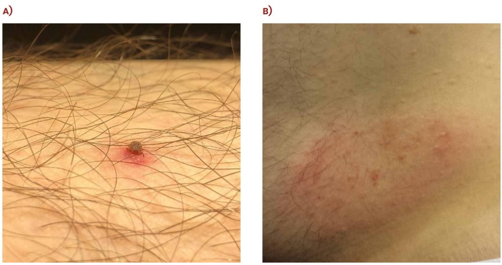
Bites from nymphal lone star ticks. A) uncomplicated bite in current study, and B) itchy Lyme disease like rash occurring after bite on the same author a year earlier. Reproduced under a Creative Commons Attribution License CC-BY 4.0 with the permission of J Goddard, JP Wyattt, https://doi.org/10.7759/cureus.29865.
New Insights
Of course, the straightforward recovery was good news for our dedicated researcher. But despite the frequency of human encounters with ticks, very little had previously been published on the progression of an uncomplicated tick bite, detailing the progress from the time of tick removal to lesion healing.
The medical assessment over 30 days, accompanied by photographic evidence, is likely the first account in the medical literature of the healing trajectory of an uncomplicated tick bite.
Dr Goddard and Dr Wyatt detail how small amounts of localised bleeding ensued following the initial bite of the tick and insertion of the hypostome. Skin irritation was greatest for the first four days, after which it subsided progressively over the 30 days. As the tick on Dr Goddard was removed promptly, the researchers argue that the development of the lesion in the following days was due to the saliva of the tick encountered at the initial bite.
The important message from this fascinating case study is that, even without complications, ticks may create skin lesions that are observable for at least 30 days. We should note that if a tick is not removed, as ensured in Dr Goddard’s case, it will continue to feed on the blood for several days (and, in doing so, rapidly increase in size). The resolution trajectories from tick bites in more complicated scenarios require further examination.
SHARE
DOWNLOAD E-BOOK
REFERENCE
https://doi.org/10.33548/SCIENTIA1015
MEET THE RESEARCHER

Dr Jerome Goddard
Mississippi State University
Mississippi State, MS
USA
Dr Jerome Goddard obtained his PhD in Medical Entomology from Mississippi State University in 1984. Directly afterwards, he served in the United States Air Force as a Captain and Medical Entomologist. In 1989, Dr Goddard was appointed State Medical Entomologist for the Mississippi State Department of Health, and in 2008, he took up his current position of Extension Professor at Mississippi State University. Dr Goddard has published over 200 scientific papers and 14 books. One of his medical textbooks, The Goddard Guide to Arthropods of Medical Importance, is in its seventh edition and was awarded ‘Highly Commended’ in the Public Health Category of the British Medical Association’s Best Medical Book of the Year in 2013. Dr Goddard has been a valuable asset to the entomological community throughout his esteemed career, as evidenced by his research, teaching and expert consultation services.
CONTACT
jgoddard@entomology.msstate.edu
W: https://www.biochemistry.msstate.edu/associate.php?id=10
KEY COLLABORATORS
Dr Julie P Wyatt, MD, Wyatt Dermatology Clinic
FURTHER READING
J Goddard, JP Wyatt, The Evolution of a Tick Bite Lesion, Cureus, 2023, 14(10), e29865. DOI: https://doi.org/10.7759/cureus.29865

REPUBLISH OUR ARTICLES
We encourage all formats of sharing and republishing of our articles. Whether you want to host on your website, publication or blog, we welcome this. Find out more
Creative Commons Licence (CC BY 4.0)
This work is licensed under a Creative Commons Attribution 4.0 International License. 
What does this mean?
Share: You can copy and redistribute the material in any medium or format
Adapt: You can change, and build upon the material for any purpose, even commercially.
Credit: You must give appropriate credit, provide a link to the license, and indicate if changes were made.
SUBSCRIBE NOW
Follow Us
MORE ARTICLES YOU MAY LIKE

The Economic Case for Prevention: Michigan Research Team Shows Diabetes Prevention Programmes Pay Dividends
For decades, healthcare systems have focused primarily on treating diseases rather than preventing them. Now, groundbreaking research from the University of Michigan demonstrates that investing in prevention – particularly for type 2 diabetes – can improve health outcomes and significantly reduce costs. Their comprehensive studies provide compelling evidence that could reshape how we approach chronic diseases.

Fostering Innovation in Healthcare Through Collaborative Learning and Research
Healthcare systems worldwide face mounting pressures from rising costs, ageing populations, and increasingly complex patient needs. To address these challenges, researchers at the All-Wales Intensive Learning Academy for Innovation in Health and Social Care at Swansea University are exploring innovative approaches that deliver better value in healthcare provision. The team led by Dr Daniel Rees and Dr Roderick Thomas are leading efforts to develop new models of collaborative learning and research that bring together healthcare providers, industry partners, and academic institutions to drive innovation in health and social care.

Epigenetic Mysteries Unravelled: The Zinc-Finger Proteins
Exploring the complex mechanisms of cell development processes and DNA structure is critical to understanding how certain diseases, such as cancer, can arise. Professor Danny Reinberg and Dr Havva Ortabozkoyun from the University of Miami in Florida, USA, work to reveal the epigenetic mechanisms at play during cell division and development and, in turn, disease processes. Together, they are discovering new protein molecules involved in genome organisation, deepening our understanding of how cancers and other related conditions can develop.

International Isocyanate Institute | TDI-induced Asthma: Reanalysing Data to Find Hidden Trends
Even if you’ve never heard of them, you’ve used polyurethanes. Producing them requires toluene diisocyanates, which may/can induce asthma when inhaled. A 5-year study claimed to conclude that cumulative TDI exposure over time was indicative of asthma incidence. However, a reanalysis by a team at the International Isocyanate Institute points the finger instead at the frequency of unprotected high-exposure events, like accidental spills or plant maintenance. This finding guides the way for future advances in worker safety.