
Professor Christine Larson – Understanding Brain Function, Cognition, and Emotion in Psychopathology

Mental health conditions such as anxiety disorders significantly impact on the quality of life of sufferers, their physical health and psycho-social functioning. Given the high prevalence and extent of impairment inflicted on affected individuals, the economic cost to public health is substantial. Professor Christine Larson at the University of Wisconsin-Milwaukee, USA, seeks to identify new and more effective targets for intervention by better understanding the relationship between brain function, cognitive processing, and emotion.
From Survival to Pathology
From an evolutionary perspective, our survival depends on the rapid identification of threats in the environment. Known as the fear response, this detection of threat occurs quickly and automatically without the need for detailed processing of the immediate situation. In a life or death situation, such as being faced by a predator, it is critical to react quickly and decisively to maximise the potential for successful ‘flight or fight’.
In modern-day life, however, this mechanism may malfunction leading to psychopathological states, such as anxiety disorders. While some psychological unease and discomfort is an inherent experience at some periods through our lives, in more severe and extreme forms it becomes a pathological reaction, significantly impacting on the quality of life of sufferers, their physical health and also their psycho-social functioning. Symptoms of clinical anxiety, such as generalised anxiety disorder, include recurrent, intense worries and fears in relation to everyday situations accompanied by physiological responses such as shaking, sweating and difficulty in breathing.
Understanding the links between brain function, cognition, and emotion is fundamental to better understanding and treating common clinical disorders.
Fear and the Amygdala: Links to Anxiety
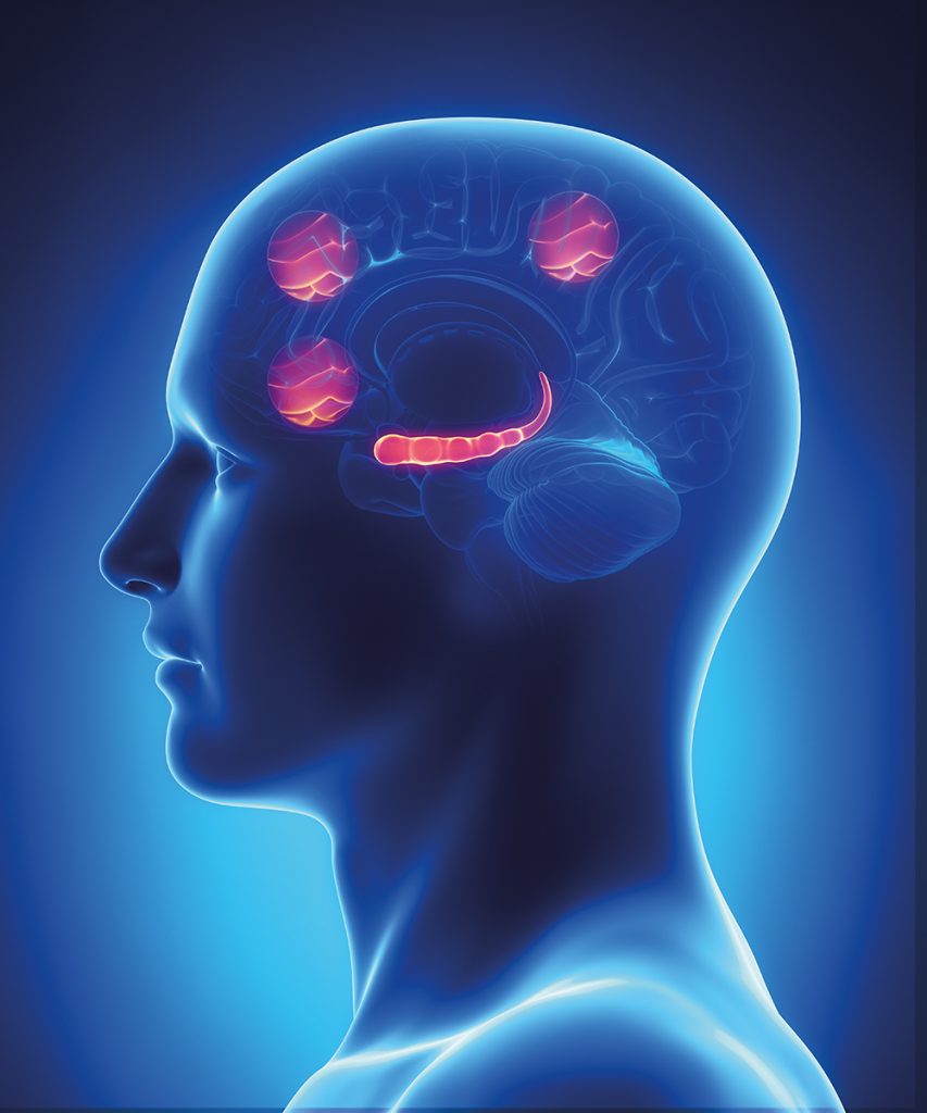
A large and established body of research implicates the amygdala, a small almond-shaped mass of cells in the brain, as being critical to fear responses. Laboratory research in animals has shown that lesions to the amygdala inhibit their usual fear responses to threat stimuli. In humans, neuroimaging studies of the brain have shown increased activation in the amygdala when participants are viewing negative stimuli, such as fearful faces.
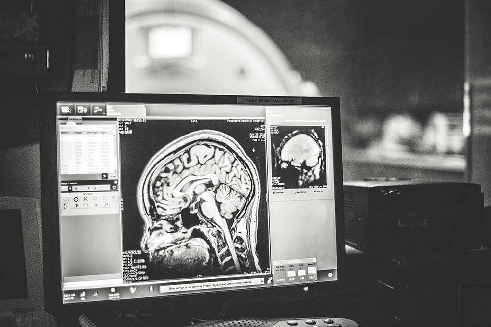
Professor Christine Larson and her team at the University of Wisconsin-Milwaukee, USA, extended this work by using functional magnetic resonance imaging (fMRI) to explore the underlying relationship between dispositional anxiety and amygdala activity, and the links with performance on an emotional working memory task. Working memory is the mental process through which we hold information in mind for immediate use (such as when we rehearse a telephone number). Working memory has only limited capacity, and as such, there is only so much information we can hold in our immediate mind at any one time).
In the emotional working memory task used by Professor Larson and her team, threatening faces were used to assess the ability of participants to remove their working memory bias away from fearful stimuli in order to effectively hold neutral stimuli in working memory. The team found that heightened amygdala activity as assessed using fMRI was associated with poorer performance on the emotional working memory task, and that this effect was greater in those participants with higher dispositional anxiety, which was assessed using a self-report measure. These findings are important because dispositional anxiety is associated with increased risk for later developing clinical anxiety, and this seems to be driven by increased amygdala activity in response to threat.
Brain Networks Underlying Anxiety
Much work in laboratory animals has focussed on identifying the brain networks between different parts of the brain associated the learning of fear responses, and the unlearning of these –also known as fear extinction. As you might expect, the amygdala plays an important role, as do the parts of the brain associated with learning and memory, such as the hippocampus. Research in animals has shown that successful fear extinction depends on plastic changes in connectivity between these regions that happened during extinction learning. Much less work, however, has been conducted in humans so such changes in connectivity are not so well understood.
Professor Larson and her team conducted a study to address this by examining the underlying mechanisms of fear extinction in human participants. Participants underwent fMRI scanning to detect changes in connectivity between key brain regions of interest, including the amygdala and hippocampus, while completing established paradigms for the learning and extinction of fear responses. Unexpectedly, they found that participants with higher levels of dispositional anxiety did not demonstrate weaker fear inhibition connectivity pathways in the brain, but rather, a strengthening of the fear expression pathways during extinction training. In other words, for individuals with dispositional anxiety, a maladaptive strengthening of fear responding meant that attempts to reduce or extinguish this were unsuccessful.
‘Our long-term goal is to characterise the neurobiology of cognitive-affective factors that confer risk for long-term distress in the aftermath of a trauma, ultimately with the goal of identifying individuals in most need of early intervention.’
Phobias, Fear, and the Amygdala
In a separate line of research, Professor Larson and her team explored whether phobic reactions may also depend on amygdala activity. A phobia is a specific type of anxiety disorder in which individuals have an intense and irrational fear of objects, places, or situations. Common examples of these include heights, spiders, and snakes. In the UK for example, spiders are rarely venomous to the extent that they could harm a human, but spider phobia is extremely common. Although an individual with a specific phobia may know that their fear is irrational, they are unable to inhibit their excessive fear reactions associated with it, with a debilitating effect on their daily lives.
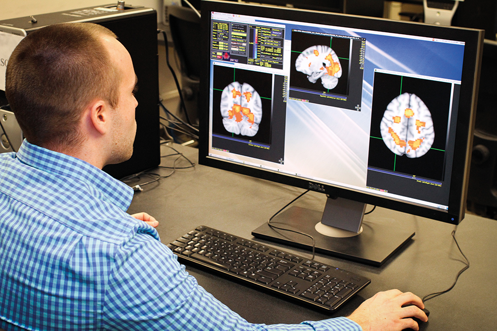
Using fMRI to assess amygdala activity, Professor Larson and her team compared the reactions of female participants who were either spider-phobic or non-phobic to pictures that contained either spider stimuli, negative stimuli, or neutral stimuli. By closely examining the time-course of amygdala activity in response to the pictures, they identified that the amygdala of spider-phobic individuals responded much more quickly to pictures of spiders than to the other types of pictures and also when compared to the non-phobic individuals. The researchers suggest that individuals with phobias are ‘over tuned’ to respond to information related to the target of their specific phobia.
The Underlying Brain Networks of a Personality Trait
Harm avoidance is a personality trait in which individuals tend to avoid specific situations that they perceive as being dangerous (rather than those that are objectively considered dangerous). An example might that of being overly concerned and worried about being in an unfamiliar situation that others feel comfortable about.
Personality traits, such as harm avoidance, reflect relatively stable characteristics associated with an individual over the course of their lifetime and for this reason, are likely to have established brain connectivity networks. However, much less is known about such brain connectivity networks than for diagnosable disorders such as anxiety and depression, which are likely to fluctuate in presence and severity over time.
Professor Larson and her team used fMRI to explore the connectivity between brain networks in a sample of young and healthy participants who had completed questionnaire measures of anxiety and harm avoidance and who were simply resting in the scanner (rather than completing any form of specific task as in their earlier studies). Previous researchers had identified disruption to several brain connectivity networks in depression and anxiety. To their surprise, Professor Larson and her team did not replicate these findings in terms of participants’ dispositional anxiety, and there was no apparent relationship between anxiety and harm avoidance, suggesting that these are separate psychological constructs. However, they demonstrated that harm avoidance is associated with disrupted functional connections in key networks which the researchers suggest reflect difficulties in the ability of individuals to distract their attention from feared objects or situations.
Building on Progress
The work already conducted by Professor Larson and her team has shone new light on how anxiety disorders and specific personality traits are underpinned by specific brain activity and connectivity between regions. Understanding these underlying processes and how they relate to cognitive processing and emotion is critical to the development of novel and much-needed interventions for mental health difficulties.
Professor Larson and her team are taking all this work forward. They now plan to further unravel the individual differences in both anxiety and vulnerability to anxiety. They also plan to identify the genetics associated with the brain function correlates of key cognitive processes such as learning and extinction of fear responses in anxiety that they so elegantly demonstrated in their previous research.
Finally, Professor Larson and her team are also turning their attention to posttraumatic stress disorder (PTSD), a psychiatric disorder that for some individuals, develops in the aftermath of trauma. PTSD is associated with significant distress and impairment, high rates of co-morbidity with other disorders such as anxiety and depression, as well as risk of suicidality. Exposure to trauma is extremely common and not everyone exposed to trauma develops PTSD. What is critical is that we don’t know what factors in the immediate post-trauma period predict the development of later PTSD for an individual.
Professor Larson and her team now plan to utilise their extensive knowledge base and experimental methodologies to better understand which individuals are at risk of developing PTSD after trauma. In a concluding note, Professor Larson explains, ‘Our long-term goal is to characterise the neurobiology of cognitive-affective factors that confer risk for long-term distress in the aftermath of a trauma, ultimately with the goal of identifying individuals in most need of early intervention.’
Reference
https://doi.org/10.33548/SCIENTIA406
Meet the researcher

Professor Christine Larson
Department of Psychology
University of Wisconsin-Milwaukee
Milwaukee, WI
USA
Professor Christine Larson completed her PhD in clinical psychology at the University of Wisconsin-Madison in 2003. Following a position at Michigan State University, Professor Larson returned to Wisconsin at the University of Wisconsin-Milwaukee in 2007 in the role of Assistant Professor. Here, she rapidly rose through the academic ranks and is now Professor of Psychology as well as Director of Clinical Training. With her research group, she explores the cognitive-affective processes associated with psychopathology using multimodal neuroimaging, electrophysiological, behavioural, and molecular genetic techniques. Professor Larson has attracted substantial funding to support her work and has published more than 60 peer-reviewed papers and three invited book chapters to date. She has received numerous honours and awards, and in 2016 was elected Secretary of the Society for Psychophysiological Research.
CONTACT
CURRENT FUNDING
National Institute of Mental Health
National Institute of Health
Medical College of Wisconsin
Marquette University Innovation Fund
FURTHER READING
EL Belleau, WS Pedersen, TA Miskovich, FJ Helmstetter, CL Larson, Cortico-limbic connectivity changes following fear extinction and relationships with trait anxiety, Social Cognitive and Affective Neuroscience, 2018, 1037–1046.
AA Huggins, EL Belleau, TA Miskovich, WS Pedersen, CL Larson, Moderating effects of harm avoidance on resting-state functional connectivity of the anterior insula, Frontiers in Human Neuroscience, 2018, 12, Article 447.
DM Stout, AJ Shackman, WS Pedersen, TA Miskovich, CL Larson, Neural circuitry governing anxious individuals’ mis-allocation of working memory to threat, Scientific Reports, 2017, 7, 8742.
EL Belleau, L Taubitz, CL Larson, Imbalance of default mode and regulatory networks during externally-focused processing in depression. Social, Cognitive, and Affective Neuroscience, 2015, 10, 744–751.
CL Larson, HS Schaefer, GJ Siegle, CAB Jackson, MH Anderle, RJ Davidson, Fear is fast in phobic individuals: amygdala activation in response to fear-relevant stimuli, Biological Psychiatry, 2006, 60, 410–417.


Creative Commons Licence
(CC BY 4.0)
This work is licensed under a Creative Commons Attribution 4.0 International License. 
What does this mean?
Share: You can copy and redistribute the material in any medium or format
Adapt: You can change, and build upon the material for any purpose, even commercially.
Credit: You must give appropriate credit, provide a link to the license, and indicate if changes were made.
More articles you may like

Dr Haydee Laza | Identifying Aphidresistance to Protect Future Crops
Insect pests, such as aphids, are capable of decimating crop yields, leading to severe economic loss. Some plants, however, have developed traits that make them resistant to such pests. Identifying these traits in plants, however, is not simple. Dr. Haydee Laza from Texas Tech University has worked with colleagues to identify several sorghum aphidresistant leaf structure traits in sorghums. These traits can be used as the basis for developing future screening methods for identifying aphid resistance in sorghums, subsequently protecting future crop yields.

Dr Rhett Martin | Governing Nature at the Edge of Collapse
For millions of men, ageing brings with it a set of frustrating and often disruptive urinary symptoms. These symptoms, caused by benign prostatic hyperplasia, or BPH, can affect sleep, confidence, and overall quality of life. Traditionally, treatment follows a familiar path. Patients begin with medications, often for years, and may eventually progress to surgery if symptoms worsen. Yet this pathway is not without its drawbacks. Medications can cause side effects, while surgery carries risks and recovery time. In recent years, a minimally invasive interventional radiology procedure called prostate artery embolisation, or PAE, has begun to challenge this traditional model. At the forefront of this shift is a collaborative research group, led by Dr. Nicholas Brown of the University of Queensland, whose series of P-EASY studies has explored whether PAE could transform how BPH is treated, particularly at earlier stages.

Assoc Prof. Nicholas Brown | Rethinking Prostate Care: A New Frontier in Treating Benign Prostatic Hyperplasia
For millions of men, ageing brings with it a set of frustrating and often disruptive urinary symptoms. These symptoms, caused by benign prostatic hyperplasia, or BPH, can affect sleep, confidence, and overall quality of life. Traditionally, treatment follows a familiar path. Patients begin with medications, often for years, and may eventually progress to surgery if symptoms worsen. Yet this pathway is not without its drawbacks. Medications can cause side effects, while surgery carries risks and recovery time. In recent years, a minimally invasive interventional radiology procedure called prostate artery embolisation, or PAE, has begun to challenge this traditional model. At the forefront of this shift is a collaborative research group, led by Dr. Nicholas Brown of the University of Queensland, whose series of P-EASY studies has explored whether PAE could transform how BPH is treated, particularly at earlier stages.

Jean Lycke | Addressing Unmet Medical Needs in Mucosal Disease: A Close-to-Market Innovation Approach
Recurrent Aphthous Stomatitis (RAS) is an oral condition characterized by one or several painful mucosal ulcers. RAS affects a large proportion of the population and has a point prevalence of approximately 2–3%, daily. The etiology remains unknown, and there is currently no curative treatment. Most patients experience recurring episodes over time, with each episode typically lasting up to a week. Here, we describe the development of a mucoadhesive patch which, when applied over a RAS ulcer, provides rapid pain relief. The patch is easy for patients to apply when symptoms begin and has the potential to be used as an over-the-counter product. The development of the Mucocort mucoadhesive patch is an example of a Close-to-Market innovation strategy that embraces simplicity within a complex healthcare system. By simplifying the product concept, the team has reduced the number of regulatory steps required before market approval. This MedTech/Pharma innovation model, known as the “4R” framework – Re-purposing, Re-formulation, Re-positioning, and Re-patenting – has guided the program from concept to commercialization. In addition to the biodegradable mucoadhesive patch developed for RAS ulcers, the team is extending the innovation concept to a mucoadhesive gel formulation for the prevention and treatment of chemotherapy-induced mucositis. This gel-based program is being commercialized separately through MucoShield.