
Professor Paul Lecoq – TICAL Aims for Paradigm Shift in PET Imaging
In an ongoing effort to increase the accuracy and sensitivity of current PET (positron emission tomography) scanners, Prof Paul Lecoq and his team at CERN research various elements of this particular imaging technique. Inspired by particle physics detectors, the team is making ground-breaking modifications to current PET technology, which will help physicians make more precise diagnoses, implement more accurate treatment plans and increase patient survival rates for a multitude of conditions. From cancer and neurodegenerative conditions through to osteoarthritis and diabetes, all will see improved diagnoses and treatment regimens for an increased number of diseases and patients.
PET as a Tool in the Fight Against Disease
Medical scanning technology has progressed swiftly over recent times. The implementation of various modalities such as magnetic resonance imaging (MRI), computed tomography (CT) and positron emission tomography (PET) has seen the medical sector enriched with many imaging techniques. These techniques have become essential tools in the early detection of many medical conditions, such as cancer, allowing for swift treatment and improved survival rates.
Because of its ability to study biochemical functions within the body, PET is often able to detect disease before the resulting changes in anatomy become apparent, making it a highly effective tool in diagnosing many diseases. However, one significant limitation of PET is that it exposes the body to radioactivity, making it unsuitable for use on children or pregnant women. Therefore, it is essential for research to improve the sensitivity of the technique so that radioactive doses can be reduced.
PET scanning works initially by injecting the patient with a substance called a radiotracer that homes in on the affected area. A specific radiotracer is selected for the patient depending on their disease and the body area to be scanned. In the case of cancer, this substance, similar to glucose, does not metabolise and accumulates in cancer cells, as they require more energy than normal cells for their fast and uncontrolled multiplication. Each radiotracer contains a radioactive element that constantly releases positively charged sub-atomic particles called positrons (the antimatter equivalent of electrons). When a positron meets an electron, both annihilate, releasing two oppositely directed gamma-ray photons. It is the role of the PET scanner to trace the path of these photons back to where they originated to isolate the disease centre.

Photonic crystal: hexagonally close-packed array of nano-sized cones
Download
Listen
Reference
https://doi.org/10.26320/SCIENTIA36
Share
The detectors contained within each PET scanner are in a 360-degree configuration surrounding the patient, so the constant flow of gamma rays from the annihilation events builds up a 3D picture of the diseased area. Physicians can then use these high-quality images to deliver treatments to their patients.
The major benefit of PET scanners over other imaging modalities is that it can directly measure complex metabolic processes, offering doctors the confidence to target highly critical molecular pathways of the activity of cells in different organs of the body, such as the brain or heart. While MRI can sometimes indirectly measure metabolic processes, it does so with poor sensitivity, at ‘millimolar’ or ‘micromolar’ concentrations. By comparison, current PET technology can analyse biochemical processes at the ‘picomolar’ level, making it over a million times more sensitive than MRI.
Improving Current PET Technology
To increase PET’s sensitivity even further, researchers are attempting to achieve more precise timing information of the detected gamma-ray photons. This will allow a more detailed picture of the disease site to be obtained, through strongly enhancing the signal to noise ratio of the image. The socalled ‘coincidence time resolution’ (CTR) of a PET detector is the measure of how precise the timing information is. In current PET scanners, the CTR is approximately 500 picoseconds (or 500 trillionths of a second), which corresponds to an imprecision of 7.5 cm in the localisation of the event at the origin of the emission of the two gamma rays along the line of response (the line between the two corresponding detectors).
When photons are registered by detectors in a PET scanner, they are organised into time intervals, or time-bins. However, if the timing resolution is too low, uncorrelated particles are also collected within each interval, creating false readings. These spurious events create additional system noise and can corrupt potential image information. Fast CTR is therefore vitally important for medical imaging.

Although 500 picoseconds might seem fast in everyday terms, time resolution is an area that Professor Lecoq and his team in the Time Imaging CALorimeter project (TICAL) intend to improve. As Professor Lecoq highlights in a recent IEEE journal submission, this ambitious upgrade down to about 10 picoseconds is physically attainable, and will lead to vast improvements in image accuracy and signalto-noise ratios. This is critical for improving the PET technique, as increased sensitivity will allow physicians to gather higher quality information faster and to reduce radiotracer doses, allowing in particular young children and even pregnant women to benefit from using PET.
Under a five-year European Research Council project, Professor Lecoq and his multinational team have been tasked to develop various aspects of the PET detection chain, from the optical materials responsible for detecting gamma rays, to the electronics that reads the converted electrical signals. Many aspects of the detection chain are responsible for timing delays, thus reducing the time resolution. For this reason, the entire five-year TICAL project has been broken down into several individual areas for investigation, with the primary purpose of reducing the time resolution to 10 picoseconds. This remarkable level of time resolution would be comparable to those achieved by particle physics detectors, such as those found at CERN.
Scintillation: Converting Light to Electrical Current
One key part of the detection process, where the magic happens, is the conversion of gamma rays into electrical signals. This is partly achieved by optical materials called ‘scintillators’ that are imbedded within the surrounding scanner (these materials are also used in particle physics detectors and have played a significant role in the discovery of the Higgs boson at CERN). When gamma-ray photons are absorbed by the atoms in a scintillator crystal, they become excited, causing them to emit light in a process known as fluorescence. This fluorescent light is then passed through a silicon-based photomultiplier (SiPM) where it is converted into an electrical current. Because scintillators play a pivotal role in the signal-conversion process, maximising how efficiently they can transport light is an important approach to improving PET imaging.
Scintillation materials come in many forms, from organic liquids, such as benzene, to gases and inorganic crystals. Because of its good performance and flexibility, Professor Lecoq and his team are concentrating their efforts on developing a metamaterial, combining the necessary high density (to allow gamma rays to efficiently interact) and good scintillation properties of lutetium oxyothosilicate (LSO) with the ultrafast light emission of nano-scintillators made from zinc oxide or cadmium selenide. To date, the team’s various techniques to improve the light output (amount of light released) from these LSO crystals have progressed well. This has primarily involved coating the scintillator’s exit face – where light leaves to enter the SiPM – using nanoimprinting technology. These so-called ‘photonic crystals’ can be imprinted on several materials (preferably with a high refraction index), such as silicon nitride and titanium dioxide, onto the scintillator’s surface.
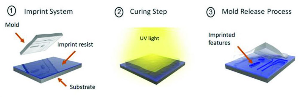
Schematic of the nanoimprinting process used for fabricating the photonic crystals
Light bouncing around within scintillator crystals has been shown to be a significant area of time delay that inevitably reduces the light output efficiency. To aid this process, the TICAL team has developed an integrated computer simulation program. By integrating several job specific programs into one modelling platform, the researchers have been able to scrutinise important system variables such as light-ray direction and photonic crystal configurations. This has enabled the team to model specific photonic crystal coating geometries to maximise light output. The successful use of the team’s innovative software scheme has already helped produce positive results, and in total, the team has not only increased the light efficiency output by 150%, but also significantly reduced the time it takes for the signal to be transported.
In conjunction with the advances made in the modelling software, the team has organised a collaboration with the Massachusetts Institute of Technology (MIT) and the companies Radiation Monitoring Devices and aBeam Technologies in USA, to develop a scalable and cost-effective production method for highly efficient photonic crystals. This has primarily involved engineering the individual photonic crystals into nano-sized cones.
As an additional research area, the team initiated the ULTIMA project to run in parallel with the TICAL project. This proof-of-concept project explores the use of engineered photonic crystals to boost the light output. When using their nanoimprinted photonic crystals, Professor Lecoq and the team have so far reported an increase in scintillated light output of greater than 40% and an increased energy resolution of up to 40% compared to currently-used non-coated scintillators. The team also highlights that, for commercial purposes, the process of nanoimprinting has been shown to be highly scalable as well as cost effective. ULTIMA has placed the project on a successful trajectory, as improved light output means a far superior image quality and hence a reduced need for high radioactive tracer doses prior to the scanning process.
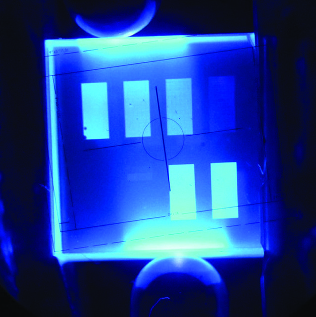
Enhanced scintillation light extraction from a UV-excited LSO crystal, on which six different photonic crystal patterns have been produced
Additional Developments and Future Applications
Under the overall management of Professor Lecoq, the entire TICAL project has been a coordinated effort between five individual teams of scientists from Fermilab in the US, the University of Bologna and CERN itself. All have been tasked to investigate additional areas of the detection chain with the ultimate goal of creating a highly accurate detector and reducing coincidence timing resolution to their target of 10 picoseconds.
This research will also have a multifunctional use, in that the team’s detection materials will eventually also be integrated into future high-luminosity particle accelerators. Detectors (specifically called calorimeters) are used in particle physics to measure the energy of particle showers in atom smashing experiments. It is predicted that the research carried out by the TICAL and ULTIMA teams will be the initial steps on a path that will trigger a cascade of further research and development projects into PET imaging technologies. The hope is they will eventually crossover from academic research into everyday use, particularly within the medical sector.
Also, the researchers are working towards new advances in silicon-based photomultipliers. Their new method, based on a silicon strip arrangement, backed up with improved readout capabilities, will help to bring the team’s timing resolution ambitions within realistic view. The recently approved TWIST proof-of-concept project will run in parallel with TICAL and concentrate on the development of a PET module using this new photodetector technology.
PET was first introduced by David E. Kuhl of the University of Michigan in the 1950s, and has been in development under various guises ever since. However, as already mentioned, PET usage is currently restricted to the adult population. Over recent times, there have been many innovative advancements in medical imaging, from silicon based detectors used in proton beam scanners, to real-time near-infrared techniques to monitor malfunctioning lymphatic systems. Entering this pantheon of hi-tech innovation, TICAL and ULTIMA are working towards a paradigm shift in PET imaging systems, thereby widening its presently limited application. The team’s ambitious goal to reduce the timing resolution to 10 picoseconds will increase the method’s sensitivity, allowing doctors to reduce potentially harmful radiotracer doses, thus ensuring the technique becomes a truly universal diagnostic tool for future generations.
Meet the researcher

Professor Paul Lecoq
CERN
Geneva
Switzerland
Professor Paul Lecoq received his diploma in Engineering at the Ecole Polytechnique de Grenoble in 1972, under the leadership of Nobel Laureate Louis Néel. After two years of research at the Nuclear Physics Laboratory of the University of Montreal, Canada, he received his PhD in Nuclear Physics in 1974. Since then, he has worked at CERN on five major international particle physics experiments. He worked as the technical coordinator of the electromagnetic calorimeter of the CMS experiment at CERN, which played a vital role in the discovery of the Higgs boson. Prof Lecoq is also the founder of the CERN-based international Crystal-Clear collaboration, a group of 28 institutes and companies worldwide contributing to the development of scintillator science. He created the SCINT conference series in 1992, which gathers scientists who work on the fundamental aspects, production technologies and applications of scintillators. He is a member of a number of advisory committees and of international Societies, and was the promoter of the CERIMED initiative (European Center for Research in Medical Imaging, inaugurated in Marseille in 2014) in 2002 for networking physics and medicine in the field of medical imaging. In 2008, Prof Lecoq was elected a member of the European Academy of Sciences and in 2017, he was made Head of the Physics division of the Academy. In 2015, he was elevated to the status of fellow at IEEE – the world’s largest technical professional organisation for the advancement of technology.
CONTACT
E: Paul.Lecoq@cern.ch
T: (+41) 22 767 6558
W: https://www.researchgate.net/profile/Paul_Lecoq
KEY COLLABORATORS
WP1: In depth study of physics processes: Adam Para, (Fermilab), Andrea Benaglia (Univ. Milano Bicocca, CERN)
WP2: Scintillator optimization: Etiennette Auffray (CERN), Rosana Martinez Turtos, (Univ. Milano Bicocca, CERN)
WP3: Light transport and photodetection optimization: Stefan Gundacker (CERN, Univ. Milano Bicocca), Matteo Salomoni (Univ. Milano Bicocca), Rosalinde Pots (RWTH Aachen, CERN), Arno Knapitsch (CERN)
WP4: Photodetectors and fast electronics: Crispin Williams (INFN Bologna, CERN), Katayoun Doroud (CERN)
WP5: 4D Calorimeter proof of concept: Tiziano Camporesi (CERN),
Marco Lucchini (Univ. Milano Bicocca, CERN)
FUNDING
European Research Council, ERC Advanced Grant #338953
![]()