
Professor Cleo Goyvaerts | New Models to Explore Lung Cancer and Develop More Effective Treatments
Lung cancer is a complex disease that remains extremely difficult to treat. Unfortunately, the conventional methods used to study it in mice have limitations when testing potential treatments. Professor Cleo Goyvaerts and Professor Hélène Salmon from Vrije Universiteit Brussel, Belgium and Institut Curie, France, respectively, worked with colleagues to develop small 3D models of lung tumours using mouse and human cells to help develop new and more effective treatments.
The Challenges of Treating Lung Cancer
Lung cancer is a leading cause of death across the world. As a complex disease with many subtypes, it can be particularly challenging to treat because it often goes undetected until it reaches an advanced stage, making it harder to treat successfully.
Immunotherapy is a new type of cancer treatment that unleashes the body’s immune system to fight cancer. It has been very successful in treating some types of cancer, but it does not work for everyone. Researchers are working to develop new ways to make immunotherapy more effective, and one approach has been to study the tumour microenvironment (TME).
As the name suggests, the TME is the environment around a tumour. It comprises many different types of cells, including immune cells, stromal cells, and blood vessels. The TME can be very immunosuppressive, meaning that it can suppress the anti-tumour activity of the immune system.
Creating 3D Lung Cancer Models
One way to study the TME is to use 3D tumour spheroids. Spheroids are clusters of cells grown in the laboratory to mimic the structure and function of tumours. Researchers have developed new methods for generating 3D lung tumour spheroids that more closely resemble human lung tumours than traditional 2D cell culture models. These models help us better understand lung cancer and test potential treatments more effectively. By mimicking the real-world conditions of lung cancer patients, these models are instrumental in guiding research efforts toward more effective therapies, ensuring that they hold promise for actual patients.
Professor Cleo Goyvaerts and her team at Vrije Universiteit Brussel in Belgium have introduced a straightforward method for generating murine (mouse-based) and human lung tumour spheroids that mimic the multi-cellular TME. These spheroids can be used to study tumour growth, immune cell infiltration, and response to therapy.
The models were made using lung cancer cells from mice and humans engineered to glow green, making them easy to spot under the microscope. They also used special lung fibroblast cells, which provide support and structure to the tumour. Finally, different types of immune cells were added to develop a new assay to test the effectiveness of immunotherapeutic drugs on these spheroids.
Creating these 3D models was like putting together a puzzle. Professor Goyvaerts and the team had to find the right balance between the cancer cells, fibroblast cells, and immune cells. Their goal was to make models that were strong, consistent in size, and formed small clusters of cancer cells. After some experiments, the team was able to craft reliable 3D lung cancer models to drive forward their research.
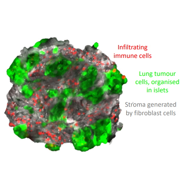
About 12 hours after mixing lung cancer cells with healthy fibroblast cells at an optimised ratio, 3D lung tumour spheroids are formed. The latter can subsequently be used to study fundamental biological processes and test novel cancer treatments.
Credit: Cleo Goyvaerts.
Pioneering Drug Combinations in 3D Tumour Models
In recent research, Professor Goyvaerts and the team confirmed the exciting potential of a new drug combination to help the body’s immune system fight lung cancer. Their spheroid model allowed the confirmation of earlier results that the combination of two existing drugs – anti-PD-1 and anti-LAG-3 – effectively kills lung cancer cells in the laboratory. This combination of drugs is now undergoing clinical trials in humans, with findings so far indicating that it is both safe and effective in the treatment of lung cancer.
‘Our uncomplicated and reproducible protocol provides a much-needed tool to advance the study and treatment of lung cancer, especially in the framework of immunotherapy.’
Pioneering Research in Tissue Communication
Collaborating with Professor Ahmed Eissa from Warwick University and Wolverhampton University (both in the UK), Professor Goyvaerts and her team have been investigating how different tissues communicate with each other without the need for animal experiments. This line of work aims to better understand how the bone marrow, representing the main source of tumour infiltrating immune cells, talks to lung tumours. More specifically, the researchers are dedicated to finding new ways to grow bone marrow cells on 3D scaffolds in the vicinity of lung tumour spheroids in the laboratory, use advanced techniques to study these cells at the single-cell level, and figure out how these interactions affect cancer outcomes and responses to treatment.
This innovative model will benefit cancer research by further reducing the need for laboratory animals. It will also improve our understanding of biomarkers for lung cancer patients, leading to more effective treatment decisions.
SHARE
DOWNLOAD E-BOOK
REFERENCE
https://doi.org/10.33548/SCIENTIA985
MEET THE RESEARCHER

Professor Cleo Goyvaerts
Laboratory for Molecular and Cellular Therapy
Department of Biomedical Sciences
Vrije Universiteit Brussel
Belgium
Professor Cleo Goyvaerts is a prominent figure in the field of cancer immunotherapy. She completed her PhD in 2015 at Vrije Universiteit Brussel (VUB) on antitumor vaccination. She then completed postdoctoral research both at VUB and Mount Sinai Icahn School of Medicine New York. Returning to VUB in 2018, Dr Goyvaerts was appointed Assistant Professor. She is also a VUB Young Leaders Academy Ambassador for EUTOPIA, a European University Alliance backed by the European Commission. Professsor Goyvaerts has earned considerable recognition, including the International Association for the Study of Lung Cancer Young Investigator Award in 2020, for her contributions to cancer immunotherapy. She is currently leading grant-supported research projects exploring bone marrow-lung cancer interactions and T-cell-mediated killing-resistant mechanisms in lung cancer. Beyond her scientific achievements, Professor Goyvaerts is a dedicated science communicator, bridging the gap between cancer research and society.
CONTACT
W: researchportal.vub.be/en/persons/cleo-goyvaerts
LinkedIn: www.linkedin.com/in/cleogoyvaerts/
KEY COLLABORATORS
- Hélène Salmon, Institut Curie, France
- Ahmed Eissa, Warwick University and Wolverhampton
University, UK - Kirsten De Ridder, Vrije Universiteit Brussel, Belgium
- Robin Maximilian Awad, Vrije Universiteit Brussel, Belgium
- Hannelore Ceuppens, Vrije Universiteit Brussel, Belgium
- Navpreet Tung, The Tisch Cancer Institute, USA
- Jan-Timon Werle, Institut Curie, France
- Léa Karpf, The Tisch Cancer Institute, USA
- Annie Bernier, Institut Curie, France
FUNDING
Fonds Wetenschappelijk Onderzoek
Kom op Tegen Kanker
Belgian American Educational Foundation
Wetenschappelijk Fonds Willy Gepts of the UZ Brussel
Belgian Council Laboratory Animal Science
Association Pour la Recherche sur le Cancer
Marie Skłodowska-Curie Actions
FURTHER READING
K De Ridder, N Tung, J Werle, et al., Novel 3D Lung Tumor Spheroids for Oncoimmunological Assays. Advanced NanoBiomed Research, 2022, 2, 2100124. DOI: https://doi.org/10.1002/anbr.202100124
![]()
REPUBLISH OUR ARTICLES
We encourage all formats of sharing and republishing of our articles. Whether you want to host on your website, publication or blog, we welcome this. Find out more
Creative Commons Licence (CC BY 4.0)
This work is licensed under a Creative Commons Attribution 4.0 International License. 
What does this mean?
Share: You can copy and redistribute the material in any medium or format
Adapt: You can change, and build upon the material for any purpose, even commercially.
Credit: You must give appropriate credit, provide a link to the license, and indicate if changes were made.
SUBSCRIBE NOW
Follow Us
MORE ARTICLES YOU MAY LIKE

Dr JoLee Sasakamoose – Dr Mamata Pandey | Empowering Indigenous Health: The Indigenous Wellness Research Collaborative in Saskatchewan
The Indigenous Wellness Research Collaborative is a transformative alliance dedicated to advancing health systems and service delivery for Indigenous communities across Saskatchewan. Founded a decade ago and co-led by Dr Mamata Pandey, a research scientist at the Saskatchewan Health Authority, and Dr JoLee Sasakamoose (M’Chigeeng First Nation), Canadian Institute of Health Research Chair in Indigenous Wellness and Health Equity at the University of Regina, their team’s work is rooted in a commitment to Indigenous leadership and community-defined wellness goals. Guided by the Cultural Responsiveness Framework, the Collaborative prioritises creating ethical spaces that serve as a middle ground for respect, reciprocity, and authentic partnerships. The team employs a strengths-based approach to health research, centering Indigenous methodologies that respect the interconnectedness of spiritual, mental, emotional, and physical well-being.

Professor Jaya Krishnan | Revolutionary Gene Therapy Helps Hearts Regenerate After Heart Attacks
Myocardial infarction, commonly termed as a heart attack, is a major cause of death and poor health worldwide. Regenerating heart tissue is an exciting and promising concept that can have significant benefits in myocardial infarctions and related diseases, but this has not yet been achieved in real-life clinical treatments. In a collaboration between Goethe University Frankfurt and Goethe University Hospital, Professor Jaya Krishnan and colleagues address this by controlling pathologic genes involved in the development of heart failure that develops after heart attacks. The researchers demonstrate a new way of treating heart disease by aiding in the division and regrowth of heart cells after a heart attack.

James J. Driscoll, MD, PhD | Immunoproteasome Activation Enhances the Recognition of Tumour Cells and Boosts Anticancer Immune Responses
The correct functioning of the human immune system depends on its ability to recognise danger, such as tumour cells, viruses, and bacteria. Scientists are learning how immunoproteasome activation can overcome the mechanisms by which cancer cells escape immune responses. Immunoproteasomes are small high molecular weight protein-degrading machines that signpost abnormal proteins made by cancer cells, directing the immune system against them. Dr James Driscoll at University Hospitals Cleveland Medical Center is using novel proprietary small molecules to selectively boost the catalytic activity of immunoproteasomes, which increases the tumour killing (or cytotoxic) effect of a group of white cells called T-cells. These findings provide a strong rationale for developing personalised therapeutics that target immunoproteasomes, for cancer and other immune-mediated conditions.

New Approaches to Treating Alzheimer’s Disease
Alzheimer’s disease is a devastating condition that strips away people’s memory, thinking, and independence. By 2050, it is expected to affect over 100 million people around the world, making it a high priority for scientific and medical research. Researchers are now exploring the potential for mechanical and light-based stimulation of the brain and nervous system to treat Alzheimer’s disease symptoms. At the University of Minho in Portugal, Francisca Monteiro is developing a PhD project supervised by a multidisciplinary set of experienced researchers, who have reviewed the evidence behind these approaches, including whole-body vibration, auditory stimulation, transcranial ultrasound stimulation, and photobiomodulation. The team aims to synthesize the evidence to support these treatments and understand what further work is needed.