
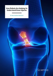
Dr Alan Barhorst | How Robots Are Helping Us Understand Knee Injuries
Knee injuries can be notoriously complex. In recent years, many studies have attempted to investigate a potential link between the geometry of the knee and the risk of injury to a ligament called the ACL (anterior cruciate ligament). Dr Alan Barhorst from the University of Lousiana at Lafayette, alongside colleague Mr Ross Wilson, enlisted the help of two robots to perform a biomechanical study of this phenomenon. Their findings provide valuable insight into our vulnerability to ACL injuries.
The Problem with Knee Injuries
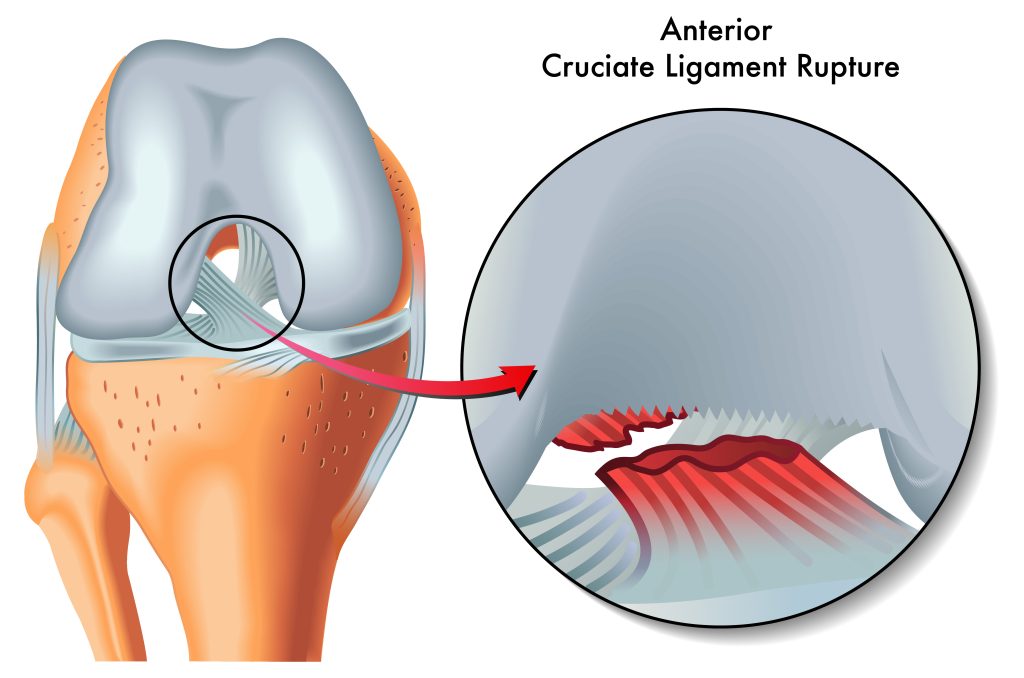
In our bodies, the ACL – known in full as the anterior cruciate ligament – provides a thick sheath of connective tissue that not only stabilises our knee but prevents our shinbone (tibia) from sliding in front of our thighbone (femur).
The ACL is a crucial area of study for several reasons. First, the American Board of Orthopaedic Surgeons reports ACL surgery to be one of the most common orthopaedic (bone or muscle) surgeries performed worldwide. Second, ACL injuries are very common in sports that involve a lot of force (e.g., jumping, contact, rapid acceleration and pivot movements). While the posterior, medial and lateral cruciate ligaments also work hard to support the knee, they can usually be rehabilitated with non-surgical treatments such as physiotherapy.
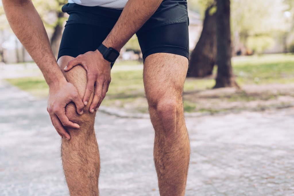
However, if you’ve ever skied, played rugby, or football, run, or are generally very active, you might have experienced ACL pain. That’s because the ACL plays a vital role in supporting and stabilising the knee in all activities, and when subjected to more stress or force than it is able to handle, it becomes injured. And that’s a problem because ACL injuries often require surgery. Dr Alan Barhorst from the University of Louisiana at Lafayette and his colleague, Mr Ross Wilson, are working to better understand how and why such injuries occur in the first place.
Taking it up a Notch
More specifically, Dr Barhorst and Mr Wilson wanted to know if a smaller-sized intercondylar notch might make us more likely to suffer from injuries to our ACL.
But what exactly is an intercondylar notch? It is called ‘intercondylar’ because it is a deep space that sits between (inter) the two condyles (or rounded ends) of our thighbone (where the thighbone attaches to the knee). It stabilises the knee and also provides the attachment site for the ACL and posterior cruciate ligaments. These ligaments play a vital role in connecting our thighbone to our shinbone.
Dr Barhorst and Mr Wilson looked specifically at whether having a smaller intercondylar notch would be more likely to cause a lateral impingement (causing a transverse load) of the ACL, and whether any observed impingement could be a cause of injury to the ACL.
The Robots Begin
To explore this question, the researchers utilised two robotic manipulators. These robots directed the knee joints of multiple cadavers (corpses), in order to simulate a range of complex biomechanical movements. The use of a two-robot system was necessary because it allowed observation of how the femur and tibia work together across a vast range of movements.
The three female and three male cadavers used were non-osteoarthritic (i.e., without arthritis), and under the age of seventy years. Biomechanical testing allowed Dr Barhorst and Mr Wilson to observe the rate of impingement in these knees, and to also observe if these impingements caused injuries. The experimental data pointed to impingements in five out of the six cadaver knees but these impingements were not a cause of injuries. This suggests that such impingements may actually be a regular and normal occurrence in healthy knees.
Male and Female Differences
Interestingly, a lower elasticity was reported in the ACL of women. That could potentially mean a lower ability to withstand force and a greater potential for injury of the ACL. Dr Barhorst and Mr Wilson also observed impingement to be more frequent in females, who also exhibited smaller intercondylar notch sizes.
Future Directions and Clinical Implications
Dr Barhorst and Mr Wilson have extended our understanding of ACL injury, providing valuable experimental data. They recommended the use of larger sample sizes, continued testing and 3D anatomical measurements in future studies. Their work ultimately contributes to a fascinating field that helps us to understand, treat, and prevent, knee injuries far more effectively. And this is great news for all of the sports people and fitness fans out there who love to stay active but injury free.
SHARE
DOWNLOAD E-BOOK
REFERENCE
https://doi.org/10.33548/SCIENTIA892
MEET THE RESEARCHER
Dr Alan Andrew Barhorst
Department of Mechanical Engineering
University of Louisiana at Lafayette
Lafayette, LA
USA
Dr Barhorst graduated from Texas A&M University in 1984 and 1989 respectively, with a BS degree and then an MS degree in Mechanical Engineering. Dr Barhost went on to complete his studies at his alma mater, being awarded a PhD in Mechanical Engineering in 1991. He is currently Professor and Head of the Mechanical Engineering Department, Alumni Association & LEQSF (Louisiana Education Quality Support Fund) at the University of Louisiana at Lafayette. In addition, Dr Barhost is the inaugural co-editor of the Springer academic journal Data Enabled Discovery and Applications. Elected a Fellow of the American Society of Mechanical Engineers in 2013, Dr Barhost was later awarded a J. Tinsley Oden Faculty Fellowship at the University of Texas at Austin in 2016. His research interests include biomechanics, control system design, dynamics, design, fluid-structure interaction and healthcare engineering.
CONTACT
E: alan.barhorst@louisiana.edu
KEY COLLABORATORS
Mr Ross Wilson, Texas Tech University, TX, USA
FUNDING
Texas Tech University College of Engineering Grant
FURTHER READING
R Wilson, AA Barhorst, Intercondylar Notch Impingement of the Anterior Cruciate Ligament: A Cadaveric In Vitro Study Using Robots, Journal of Healthcare Engineering, 2018, 8698167. DOI: http://doi.org/10.1155/2018/8698167
![]()
REPUBLISH OUR ARTICLES
We encourage all formats of sharing and republishing of our articles. Whether you want to host on your website, publication or blog, we welcome this. Find out more
Creative Commons Licence (CC BY 4.0)
This work is licensed under a Creative Commons Attribution 4.0 International License. 
What does this mean?
Share: You can copy and redistribute the material in any medium or format
Adapt: You can change, and build upon the material for any purpose, even commercially.
Credit: You must give appropriate credit, provide a link to the license, and indicate if changes were made.
SUBSCRIBE NOW
Follow Us
MORE ARTICLES YOU MAY LIKE

Dr JoLee Sasakamoose – Dr Mamata Pandey | Empowering Indigenous Health: The Indigenous Wellness Research Collaborative in Saskatchewan
The Indigenous Wellness Research Collaborative is a transformative alliance dedicated to advancing health systems and service delivery for Indigenous communities across Saskatchewan. Founded a decade ago and co-led by Dr Mamata Pandey, a research scientist at the Saskatchewan Health Authority, and Dr JoLee Sasakamoose (M’Chigeeng First Nation), Canadian Institute of Health Research Chair in Indigenous Wellness and Health Equity at the University of Regina, their team’s work is rooted in a commitment to Indigenous leadership and community-defined wellness goals. Guided by the Cultural Responsiveness Framework, the Collaborative prioritises creating ethical spaces that serve as a middle ground for respect, reciprocity, and authentic partnerships. The team employs a strengths-based approach to health research, centering Indigenous methodologies that respect the interconnectedness of spiritual, mental, emotional, and physical well-being.

Professor Jaya Krishnan | Revolutionary Gene Therapy Helps Hearts Regenerate After Heart Attacks
Myocardial infarction, commonly termed as a heart attack, is a major cause of death and poor health worldwide. Regenerating heart tissue is an exciting and promising concept that can have significant benefits in myocardial infarctions and related diseases, but this has not yet been achieved in real-life clinical treatments. In a collaboration between Goethe University Frankfurt and Goethe University Hospital, Professor Jaya Krishnan and colleagues address this by controlling pathologic genes involved in the development of heart failure that develops after heart attacks. The researchers demonstrate a new way of treating heart disease by aiding in the division and regrowth of heart cells after a heart attack.

James J. Driscoll, MD, PhD | Immunoproteasome Activation Enhances the Recognition of Tumour Cells and Boosts Anticancer Immune Responses
The correct functioning of the human immune system depends on its ability to recognise danger, such as tumour cells, viruses, and bacteria. Scientists are learning how immunoproteasome activation can overcome the mechanisms by which cancer cells escape immune responses. Immunoproteasomes are small high molecular weight protein-degrading machines that signpost abnormal proteins made by cancer cells, directing the immune system against them. Dr James Driscoll at University Hospitals Cleveland Medical Center is using novel proprietary small molecules to selectively boost the catalytic activity of immunoproteasomes, which increases the tumour killing (or cytotoxic) effect of a group of white cells called T-cells. These findings provide a strong rationale for developing personalised therapeutics that target immunoproteasomes, for cancer and other immune-mediated conditions.
New Approaches to Treating Alzheimer’s Disease
Alzheimer’s disease is a devastating condition that strips away people’s memory, thinking, and independence. By 2050, it is expected to affect over 100 million people around the world, making it a high priority for scientific and medical research. Researchers are now exploring the potential for mechanical and light-based stimulation of the brain and nervous system to treat Alzheimer’s disease symptoms. At the University of Minho in Portugal, Francisca Monteiro is developing a PhD project supervised by a multidisciplinary set of experienced researchers, who have reviewed the evidence behind these approaches, including whole-body vibration, auditory stimulation, transcranial ultrasound stimulation, and photobiomodulation. The team aims to synthesize the evidence to support these treatments and understand what further work is needed.