
Dr Philip Kennedy | The Quest for Lifetime Neural Interfaces: A Review of Electrode Technologies
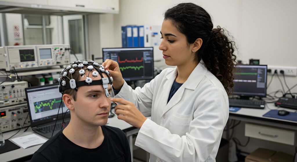
The holy grail of brain-computer interfaces is an electrode that lasts a lifetime. Dr Philip Kennedy of Neural Speech Inc. has examined the landscape of neural recording technologies, comparing their longevity and effectiveness in helping paralysed patients communicate and regain mobility.
The Challenge of Connecting Brains to Computers
For people who have severe paralysis due to conditions like motor neurone disease, brainstem stroke, or spinal cord injury, brain-computer interfaces (BCIs) offer a ray of hope. These technologies detect neural signals and translate them into commands that can control computers, robotic limbs, or speech synthesisers.
A significant challenge stands in the way of making BCIs practical, long-term solutions: the electrodes recording neural signals must remain functional for decades. Most patients would need them to work reliably throughout their remaining lifespan, potentially many decades, especially for younger patients.
Dr Philip Kennedy, a physician and researcher at Neural Speech Inc. in Georgia, USA, has focused his research on this challenge. In his comprehensive review, Dr Kennedy analyses various electrodes used in BCIs, examining their designs, advantages, limitations, and, crucially, their longevity.
Pioneering research, early milestones
Dr Kennedy began his work on neural interfaces in the 1980s, developing the Neurotrophic Electrode. This innovative approach was inspired by research showing that neurons could grow into introduced materials. In this innovative line of investigation, he confirmed that a glass cone containing nerve tissue could be successfully implanted into the brain’s cortex. When inserted, nearby neurons would extend branch-like structures into the cone. Insulated gold wires inside could then record the electrical activity of these ingrown neural connections.
The major breakthrough came when Dr Kennedy’s team discovered that these recordings could persist as long as experimental animals lived – over a year in rats and similar timeframes in monkeys. This contrasted sharply with conventional metal electrodes, which typically lost functionality over months.
In 1996, Dr Kennedy achieved a milestone by implanting his Neurotrophic Electrode in a human patient. Since then, his team has gathered extensive human data, including one remarkable case where the electrode survived for 13 years until the patient’s death with no sign of scarring. Two other patients maintained stable recordings for four years until they passed away.

How Neurotrophic Electrodes Work
When Dr Kennedy’s glass cone electrode is implanted into the brain, it causes minor intentional damage. This damage, along with growth factors placed inside the cone, triggers nearby neurons to extend neurites into the recording area. Over several weeks, these neurites become myelinated, creating stable neural pathways.
What makes this approach unique is that the electrode does not penetrate or press against the neurons it records from. Instead, the neurons grow into the recording space voluntarily, creating a natural, stable interface.
Dr Kennedy’s histological studies have confirmed this mechanism, showing healthy neural tissue growing into the electrode with no sign of scarring even after many years. This stands in contrast to conventional electrodes, which typically become surrounded by scar tissue that degrades their recording capabilities.
The original design could only record about 40 signals per electrode, fewer than some metal arrays. However, Dr Kennedy notes that a newer version developed in collaboration with Neuronexus Inc. can record 300–400 signals from the same-sized tip, potentially overcoming this limitation.
Metal-Tipped Electrodes: High Signal Count but Limited Lifespan
Metal-tipped arrays, such as the Utah Array, have dominated much of brain-computer interface research due to their ability to record from multiple neurons simultaneously. Developed by Richard Normann and colleagues in the 1980s and first implanted in humans in 2004, these electrodes feature arrays of tiny metal tines that penetrate the brain tissue. Dr Kennedy acknowledges the impressive achievements made with these electrodes. Teams using Utah Arrays have enabled paralysed patients to control robotic arms, computer cursors, and even generate synthetic speech.
However, their longevity poses a significant challenge. Dr Kennedy cites research showing that in a cohort of 55 arrays in monkeys and 40 human implantations, the average functional lifespan was just 622 days. While some lasted longer, the yield of recorded neurons typically fell from 90% to just 15% over 36 months. Dr Kennedy describes a particularly poignant case where a patient who had learned to control a robotic arm gradually lost this ability over several years, as the electrode’s signals deteriorated.
The limited lifespan stems from three main factors: micromovements between the rigid electrode and the softer brain tissue, immune reactions to the foreign material, and scarring that electrically isolates the electrode from nearby neurons.
Emerging Alternative Approaches
Dr Kennedy’s review examines several innovative approaches being developed to address the longevity challenge. The Stentrode, developed by Synchron Inc., is inserted into blood vessels near the brain’s surface. Similar to cardiac stents, the mesh-like device integrates with the blood vessel wall and can record neural signals non-invasively.
Several teams are exploring flexible and ultra-soft materials to better match the physical properties of brain tissue. The MESH electrode incorporates subcellular-sized sensors that make intimate contact with neurons and has shown promising results in mice, with signals persisting for at least a year. A further possibility lies in ‘enhanced electrodes’ that combine features of both neurotrophic and metal-tipped approaches, including hydrogel-based designs that become supple after implantation.
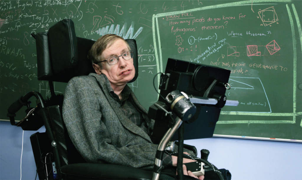
Stephen Hawking at his office at the department of applied mathematics and theoretical physics at Cambridge University in 2005. Photograph: Murdo Macleod/The Guardian
Human Impact and Ethical Considerations
Dr Kennedy emphasises the profound human implications of electrode longevity. For patients with locked-in syndrome, ALS, or spinal cord injuries, the functional lifespan of neural interfaces directly impacts their quality of life and treatment decisions.
Dr Kennedy draws particular attention to patients with ALS, who can survive for decades on ventilators – Stephen Hawking lived for 50 years after diagnosis. For these patients, a neural interface that degrades after a few years would mean returning to isolation after briefly experiencing enhanced communication abilities.
These real-world consequences raise serious ethical questions about implanting electrodes with known longevity limitations. Dr Kennedy emphasizes that patients must be fully informed about the expected lifespan of any implanted device.
The Future of Neural Interfaces
As Dr Kennedy looks to the future, he projects divergent paths for different electrode technologies. His assessment suggests that only electrodes forming intimate connections with the brain’s neural tissue will achieve the lifespans of over 50 years needed for many patients. Meanwhile, metal-tipped electrodes are likely to continue playing important roles in short-term applications. Dr Kennedy notes significant progress in regulatory approvals, with nine electrodes now approved by the US Food and Drug Administration or European regulators. This represents substantial growth from just two decades ago, when only the Neurotrophic Electrode and Utah Array had received approval.
The importance of achieving lifetime neural interfaces cannot be overstated. For locked-in patients, brainstem stroke survivors, those with ALS, or people with quadriplegia, a neural interface represents a lifeline to the world. The difference between an electrode that functions for three years versus 30 years is not merely technical – it is the difference between temporary relief and genuine life transformation. Dr Kennedy’s work, with its demonstrated 13-year functionality in one patient, represents a significant milestone toward this goal. As research continues, the most promising path forward may involve combining the best aspects of different approaches to achieve the longevity needed for truly life-changing neural interfaces.

SHARE
DOWNLOAD E-BOOK

REFERENCE
https://doi.org/10.33548/SCIENTIA1253
MEET THE RESEARCHER

Dr Philip Kennedy
Neural Speech Inc., Duluth, Georgia, USA
Dr Philip Kennedy obtained his MD from the National University of Ireland in 1972, FRCSI (General Surgery) in 1976, PhD in Neurophysiology from Northwestern University in 1983 and completed a Neurology Residency at Emory University, Atlanta, Georgia. He is the Founder and Chief Scientist of Neural Speech Inc. and has practised neurology since 1997. His pioneering work on brain-computer interfaces spans over 35 years, including the development of the Neurotrophic Electrode for direct neural recordings in humans. He has conducted six human implants, with a recent focus on speech prosthesis development for locked-in patients.
Dr Kennedy holds multiple patents and has published extensively, with over 89 peer-reviewed articles and book chapters. His work has been featured in numerous documentaries and media outlets, and he has received significant recognition, including the 2000 World Technology Award in Health and Medicine and the 2013 Neurotechnology Report’s Researcher of the Year award. His groundbreaking research continues to advance neural interface technology for restoring function in individuals with severe disabilities.
CONTACT
E: Phlkennedy@aol.com
KEY COLLABORATORS
Dinal Andreasen, Georgia Institute of Technology, Atlanta, Georgia; Marla Gearin, Neuropathologist, Department of Neurology, Emory University; Dr. Joel Cervantes, neurosurgeon, Belize City, Belize; Dr. Princewill Ehirim, Neurosurgeon, Lawsrenceville, Georgia.
FUNDING
National Institutes of Health (NINDS and NIDCD); Paralyzed Veterans of America; Dark Pulse Inc; Emory-Georgia Tech Bioengineering Research Center (31 total grants).
FURTHER READING
PR Kennedy, AJ Cervantes, Recruitment and Differential Firing Patterns of Single Units During Conditioning to a Tone in a Mute Locked-In Human, Frontiers in Human Neuroscience, 2022, 16, 864983. DOI: https://doi.org/10.3389/fnhum.2022.864983
M Gearin, PR Kennedy, Histological confirmation of myelinated neural filaments within the tip of the Neurotrophic Electrode after a decade of neural recordings, Frontiers in Human Neuroscience, 2020, 14, 111. DOI: https://doi.org/10.3389/fnhum.2020.00111
JS Brumberg, A Nieto-Castanon, PR Kennedy, FH Guenther, Brain-Computer Interfaces for Speech Communication, Speech Communication, 2010, 52(4), 367–379. DOI: https://doi.org/10.1016/j.specom.2010.01.001
Kennedy P.R., Gambrell C, Ehirim P, and Cervantes A. Advances in the development of a speech prosthesis. Book chapter in Brain-Machine Interfaces: Uses and Developments accepted 2017.
Kennedy P.R., Dinal S. Andreasen, Jess Bartels, Princewill Ehirim, E. Joe Wright, Steven Seibert, Andre Joel Cervantes. Validation of Neurotrophic Electrode long-term recordings in human cortex. Handbook of Biomedical Engineering. 2017.
Kennedy, P.R., Andreasen D.S., Bartels, J., Ehirim P., Mao H., Velliste M.,Wichmann T.,Wright, E.J. Making the lifetime connection between brain and machine for restoring and enhancing function. Proceedings in Brain Research CH.1 August 2011.
Brumberg J., Wright EJ, Andersen D, Guenther FH and Kennedy PR. Classification of intended phoneme production from chronic intracortical microelectrode recordings in speech motor cortex. Frontiers in Neuroscience 5(65)1-14, 2011.
Brumberg JS, Nieto-Castanon A, Kennedy PR, Guenther FH. Brain-Computer Interfaces for Speech Communication. Speech Commun. 2010 Apr 1;52(4):367-379.
Guenther FH, Brumberg JS, Wright EJ, Nieto-Castanon A, Tourville JA, Panko M, Law R, Siebert SA, Bartels JL, Andreasen DS, Ehirim P, Mao H, Kennedy PR. A wireless brain-machine interface for real-time speech synthesis. PLoS One. 2009 Dec 9;4(12):e8218.
![]()
REPUBLISH OUR ARTICLES
We encourage all formats of sharing and republishing of our articles. Whether you want to host on your website, publication or blog, we welcome this. Find out more
Creative Commons Licence (CC BY 4.0)
This work is licensed under a Creative Commons Attribution 4.0 International License. 
What does this mean?
Share: You can copy and redistribute the material in any medium or format
Adapt: You can change, and build upon the material for any purpose, even commercially.
Credit: You must give appropriate credit, provide a link to the license, and indicate if changes were made.
SUBSCRIBE NOW
Follow Us
MORE ARTICLES YOU MAY LIKE
Guidelines, Governance, and Gender
What are sex and gender? While biologic or physiologic differences are tremendously important, so too are the ways that women and men experience their lives and their work differently. Women often have very different experiences of health care because of bias or prejudice within the system. Equally, men get treated differently and often don’t seek health care because of the ways that gender norms encourage them to be resilient and not seek care. So, is gender an ideology? We were luck to speak with Shirin Heidari, Thomas Babor, and Jocalyn Clark, who helped us navigate the muddy waters of the current climate.

SCIENTIA’s new look: Ziggy tells all
Since 2015, we’ve partnered with over 2,500 research teams in more than 80 countries to make their work more accessible and visible — both within academia and beyond. Because we live in a world of information overload, the Scientia concept plays a significant role extending research outreach beyond traditional journal publication — in a case study, we analyse why Roustem Miftahof has chosen us more than once to translate and distribute complex scientific information to the general public.
Case study: Returning to us
Some of the researchers we feature are keen to experience what it is like to publish an article in a magazine that is not a scholarly publication. Some want the chance to see how communicators translate their publications and data in different ways. Some are keen to showcase a completed project, while others want to explain a research plan or ongoing study. Some must abide by funder requirements, while others plan to engage patients in their own treatment. Some seek a summary of research findings that is less technical than a preprint.
Other researchers, such as Dr Roustem Miftahof, have a clear goal in mind. Here, we look into the metrics of the two articles we published based on his works in 2025, and present his testimonial of the experience.
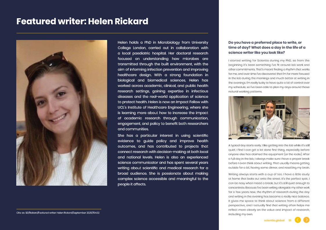
Featured writer: Helen Rickard
Helen holds a PhD in Microbiology from University College London, carried out in collaboration with a local paediatric hospital. Her doctoral research focused on understanding how microbes are transmitted through the built environment, with the aim of informing infection prevention and improving healthcare design. With a strong foundation in biological and biomedical sciences, Helen has worked across academic, clinical, and public health research settings, gaining expertise in infectious diseases and the real-world application of science to protect health. Helen is now an Impact Fellow with UCL’s Institute of Healthcare Engineering, where she is learning more about how to increase the impact of academic research through communication, engagement, and policy to benefit both researchers and communities.
She has a particular interest in using scientific evidence to guide policy and improve health outcomes, and has contributed to projects that connect research with decision-making at both local and national levels. Helen is also an experienced science communicator and has spent several years writing about scientific and medical research for a broad audience. She is passionate about making complex science accessible and meaningful to the people it affects.