
Dr Omar Islam | Portable Magnetic Resonance Imaging: An Important Innovation
Imaging technologies are vital in modern medicine and have revolutionised how clinicians make diagnoses and monitor disease progression. However, the necessary equipment – such as a scanner for magnetic resonance imaging (MRI) – is very large and expensive, requiring patients to go to the scanner rather than receiving scans as bedside care. This takes up valuable staff time and resources, and can present further risks to patients. Dr Omar Islam from Queen’s University and Drs Aditya Bharatha and Amy Lin from the University of Toronto are showing how portable MRI scanners may offer a viable alternative that benefits patients and healthcare systems.
Imaging in Medical Care
Medical imaging is crucial for accurately diagnosing illness, guiding certain types of treatment, and monitoring patient progress. Medical imaging includes technologies such as X-rays, computed tomography (CT), and magnetic resonance imaging (MRI).
MRI uses strong magnetic fields and radio waves to produce images of the body’s organs, tissues, and structures. This allows the generation of deeply detailed images, making it particularly valuable for examining the brain and organs such as the liver and heart. As well as providing detailed results, MRI is non-invasive, doesn’t require injections or incisions, and doesn’t use ionising radiation – all of which make it a safer and more comfortable option for patients.
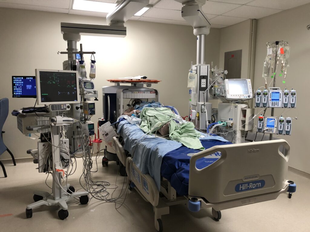
Brain imaging is especially useful in the intensive care (ICU) setting. ICU patients are typically too sick for traditional physical examinations by doctors, so MRI scans are used to provide information on brain activity, functions, and disease.
Imaging technologies such as MRI scanners are huge pieces of equipment involving a suite of rooms and a team of experts to run and analyse the tests. This means that patients requiring imaging need to be moved from the ICU. Transporting patients within the hospital is complicated and associated with substantial risks – up to 60% of transported patients may experience an adverse event, such as accidentally removing breathing tubes or equipment malfunctions. Many ICU patients will be on ventilators or life-support machines, and so will need assistance from multiple staff members (potentially diverting these limited number of staff members from taking care of other ICU patients). Researchers from Queen’s University and the University of Toronto in Canada are working to solve the issues of MRI access and hospital wait times by researching and analysing the benefits of portable MRI machines.
Portable Magnetic Resonance Imaging
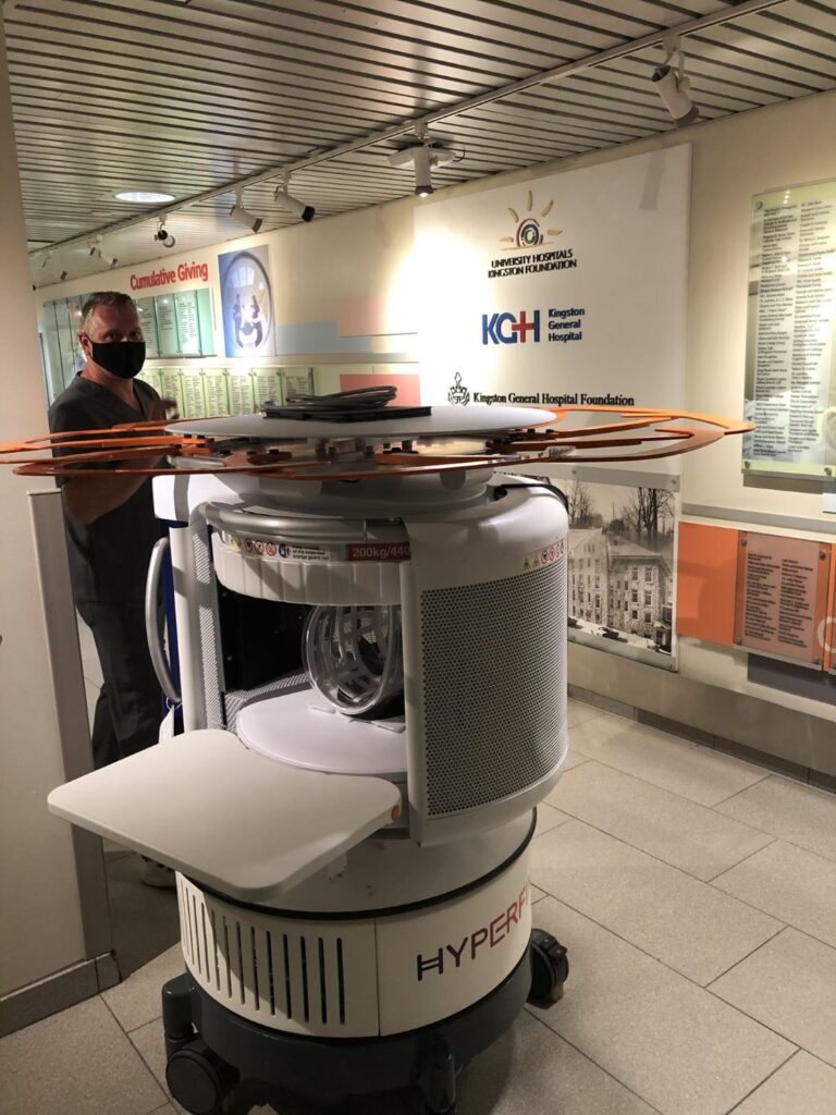
Portable MRI scanners are a recent technological innovation that allows patients to receive brain imaging without having to leave their beds or wards. A recent study confirmed that portable MRI is a safe and convenient method for ICU patients to receive imaging, with no staff or patients in the study reporting any negative events.
The HyperfineTM portable MRI scanner has been approved by the US Food and Drug Administration and Health Canada for certain standing imaging tests. Patients stay in their room throughout the scanning process, which means they can remain connected to vital breathing and blood devices, reducing the risk of equipment malfunction. This also means that doctors and nurses are able to continue with their routine work while the images are taken.
Portable MRI is known to work well for investigating certain physical changes to the brain, such as bleeds, fluid build-up, and certain types of strokes, as well as being used to direct the placement of tubes known as shunts, which allow the drainage of excessive fluid. However, portable MRI is not currently recommended for other indications such as seizures, infections, or tests where staining of the brain is required.
Investigating the potential of portable MRI in Canadian hospitals
All the requests for brain imaging for ICU patients over a whole year in two Canadian hospitals were reviewed. They analysed data from patients who needed brain imaging for conditions that may be able to be effectively and safely investigated by portable MRI. They then used patient and hospital data to compare the amount of time required for traditional imaging in radiology departments, and the number of patients that could benefit from being diverted to portable MRI. They used this information to calculate the potential time and resources that could be saved by introducing portable MRI to ICU wards.
Portable MRI Provides Significant Benefits
The team found that a significant number of brain MRI and CT scans could be performed using portable MRI instead of traditional MRI or CT technology. This would provide benefits to patient safety, staff workload, and radiology wait times.
The combined data from both hospitals showed that 24.6% of MRI and 21.0% of CT scans could be performed using portable MRI in the ICU. A typical traditional MRI scan in the hospitals in this study takes 90 minutes per ICU patient but only 30 minutes for the average outpatient. This means that if every eligible ICU patient had a portable MRI instead of being transported within the hospital for a traditional fixed MRI, the hospitals would be able to image and treat an additional 300 outpatients each year. If the eligible ICU patients who had a CT scan were able to have a portable MRI scan instead, the hospitals would be able to provide CT scans to an additional 1676 outpatients.
This work highlights the vital role portable MRI could play in the diagnosis and management of brain conditions in the ICU, and the positive implications this would have for outpatient radiology wait times. The researchers also reviewed the literature on portable MRI technology. They reported a study where portable MRI was implemented in a remote northern Canadian community which did not previously have access to traditional MRI technology. If portable MRI was implemented instead of traditional MRI and used for 50 patients a year, the hospital would save nearly eight million dollars over 5 years and improve the standard of patient care.
A New and Exciting Technology
Portable MRI is a new and exciting technology, and the benefits are not yet fully realised. An initial concern was a lack of specialised staff to operate the machines. However, in 2021, the Ontario Association of Medical Radiation Technologists announced that X-ray technologists instructed by physicians could operate any portable MRI scanner. Portable MRI technology is already progressing, and the team hopes advances will allow currently ineligible ICU patients to receive their imaging in portable MRI scanners. Validating portable MRI results against the gold standard of traditional MRI and a full-cost benefit analysis will build upon the team’s work and allow healthcare professionals and policymakers to fully understand the benefits of portable MRI scanners. The radiologists’ work is helping improve the standard of patient care and tackle the radiology wait time crisis in Canada, and further developments are to be expected in the near future.
SHARE
DOWNLOAD E-BOOK
REFERENCE
https://doi.org/10.33548/SCIENTIA1024
MEET THE RESEARCHER
Dr Omar Islam, MD FRCPC DABR
Division of Neuroradiology, Department of Diagnostic Radiology
Kingston Health Sciences Centre
Queen’s University
Kingston, Ontario
Canada
Dr Omar Islam received his medical degree from the University of Toronto and subsequently specialised as a neuroradiologist at Yale University. He is now Head of the Department of Diagnostic Radiology at Kingston Health Sciences Centre and Providence Care, and an Assistant Professor at Queen’s University in Ontario, Canada. Dr Islam sits on the Examination Board in Diagnostic Radiology for the Royal College of Physicians and Surgeons of Canada. He co-founded the Portable MRI Journal, the leading open-access source of academic information, guidelines, and policies regarding portable MRI technology. He previously served as a reviewer for the Canadian Journal of Neurological Sciences and Head & Neck. He is the Regional Imaging Lead for Cancer Care Ontario. In addition to his clinical and academic duties, Dr Islam co-founded Canadian Teleradiology Services Incorporated – one of Canada’s top Teleradiology companies.
CONTACT
L: www.linkedin.com/in/omar-islam-a898257b
KEY COLLABORATORS
Dr Amy Lin, St Michael’s Hospital, Unity Health Toronto, University of Toronto
Dr Aditya Bharatha, St Michael’s Hospital, Unity Health Toronto, University of Toronto
FUNDING
Research Grant Support, Academic Fund, Department of Diagnostic Radiology, Queen’s University
FURTHER READING
O Islam, AW Lin, A Bharatha, Potential application of ultra-low field portable MRI in the ICU to improve CT and MRI access in Canadian hospitals: a multi-center retrospective analysis, Frontiers in Neurology, 2023, 14, 1220091. DOI: https://doi.org/10.3389/fneur.2023.1220091

REPUBLISH OUR ARTICLES
We encourage all formats of sharing and republishing of our articles. Whether you want to host on your website, publication or blog, we welcome this. Find out more
Creative Commons Licence (CC BY 4.0)
This work is licensed under a Creative Commons Attribution 4.0 International License. 
What does this mean?
Share: You can copy and redistribute the material in any medium or format
Adapt: You can change, and build upon the material for any purpose, even commercially.
Credit: You must give appropriate credit, provide a link to the license, and indicate if changes were made.
SUBSCRIBE NOW
Follow Us
MORE ARTICLES YOU MAY LIKE

Jean Lycke | Addressing Unmet Medical Needs in Mucosal Disease: A Close-to-Market Innovation Approach
Recurrent Aphthous Stomatitis (RAS) is an oral condition characterized by one or several painful mucosal ulcers. RAS affects a large proportion of the population and has a point prevalence of approximately 2–3%, daily. The etiology remains unknown, and there is currently no curative treatment. Most patients experience recurring episodes over time, with each episode typically lasting up to a week. Here, we describe the development of a mucoadhesive patch which, when applied over a RAS ulcer, provides rapid pain relief. The patch is easy for patients to apply when symptoms begin and has the potential to be used as an over-the-counter product. The development of the Mucocort mucoadhesive patch is an example of a Close-to-Market innovation strategy that embraces simplicity within a complex healthcare system. By simplifying the product concept, the team has reduced the number of regulatory steps required before market approval. This MedTech/Pharma innovation model, known as the “4R” framework – Re-purposing, Re-formulation, Re-positioning, and Re-patenting – has guided the program from concept to commercialization. In addition to the biodegradable mucoadhesive patch developed for RAS ulcers, the team is extending the innovation concept to a mucoadhesive gel formulation for the prevention and treatment of chemotherapy-induced mucositis. This gel-based program is being commercialized separately through MucoShield.
The Translational Asian Agerelated Macular Degeneration Program Phase 2 (TAAP-2): Reimagining the Future of Vision Care
Age-related macular degeneration, often abbreviated as AMD, is one of the leading causes of vision loss among older adults worldwide. In Asia, where populations are ageing rapidly, its impact is particularly profound. For many, the disease quietly erodes central vision, making everyday activities such as reading, driving, and recognising faces increasingly difficult. Against this backdrop, the Translational Asian Age-related Macular Degeneration Programme, or TAAP for short, has emerged as a bold and ambitious effort to confront the disease headon. Now in its second phase, TAAP-2 represents a significant evolution in both scientific scope and clinical ambition.
Ms. Aikaterini Dritsoula | Looking Beyond Snoring: How Hidden Airway Problems Shape Children’s Sleep
For many parents, a child’s snoring may seem harmless, even endearing. Yet in some cases, it signals something more serious. Obstructive sleep apnoea is a condition in which a child’s breathing is repeatedly disrupted during sleep. These interruptions can affect growth, behaviour, and learning. Children with this condition may toss and turn at night, struggle to concentrate during the day, or show signs of hyperactivity and fatigue. Traditionally, enlarged tonsils and adenoids have been seen as the main culprits. Surgery to remove them has long been considered the standard treatment. However, research led by Consultant ENT Surgeon Ms. Aikaterini Dritsoula of The Leeds Teaching Hospitals NHS Trust invites us to look deeper. Her work suggests that the story is often more complex, especially in very young children.
Professor Neil Coffee – Professor Vincent Versace | Mapping Health Access: Using Address-Level Intelligence for Smarter Services
Accessing healthcare is a serious challenge for people living in rural and remote Australia. Large distances, sparse populations, and limited services can prevent residents from receiving care when they need it. Professors Neil Coffee and Vincent Versace at Deakin University’s Centre for Australian Research into Access (CARA) are leading research to model healthcare service access across the country, to provide new insights that can guide health planning and policy, as well as other services such as education. This work combines the curation of detailed address level residential dwellings and road network data to calculate access to service metrics (time and distance). These metrics are applied to the simulated residential dwelling population, to quantify the population with poor access to health services.