
Dr Kristin Parent – Bacteriophage Hunting: Searching for the Tiny Viruses That Kill Harmful Bacteria
Bacteriophages are tiny viruses that infect and kill bacteria but are harmless to humans. With the world facing devastating antibiotic resistance in the coming years, they may be our best hope for treating bacterial disease. There are believed to be 10^31 bacteriophages in the world, and most of them have not yet been identified. Dr Kristin Parent from Michigan State University is working on exciting, collaborative projects hunting for bacteriophages in their natural environment. She has made huge strides in elucidating the mysterious and important world of bacteriophages.
Shigella Bacteria and Bacterial Infection
Shigellosis is a nasty infection of the Shigella bacteria with over 164 million cases each year leading to 1.1 million deaths. Sadly, many of these deaths are children, as those under the age of 5 are most likely to catch a Shigella infection. This is partly due to the method of transmission – accidentally ingesting the bacteria residing in the stool of an infected person. Often this occurs in childcare settings if carers don’t thoroughly wash their hands after toilet training, for example. Swimming and drinking contaminated water or eating food by an infected person can also pass along Shigella.
The symptoms of shigellosis include fever, nausea, stomach pain and diarrhoea which often contains blood. These issues usually last for around a week, but they can result in secondary complications such as dehydration, seizures and blood infections. Whilst antibiotics are sometimes given for shigellosis, many strains are now resistant so treating the diarrhoea and consequent dehydration is often the priority until the infection runs its course.
Shigellosis is the most common form of dysentery; other types are caused by Salmonella and E. coli, among other bacteria. There are four different species of Shigella called S. boydii, S. dysenteriae, S. flexneri, and S. sonnei. S. flexneri is the most common type and is often associated with low-income countries, however, cases of S. sonnei, which tends to be found in high-income countries, are increasing. This is due to the low dose of bacteria needed to cause illness and the ever-increasing antibiotic resistance of the bacteria.
Antibiotic resistance is a huge and growing issue globally, for many different bacteria. The misuse of antibiotics in humans and animals has resulted in bacterial strains that are not killed by conventional antibiotics. When these new strains are passed on, so is the antibiotic resistance and the infections they cause are much harder to treat and mortality rates become higher. A well-known example of this is the superbug methicillin-resistant Staphylococcus aureus, better known as MRSA, a type of bacteria that is now resistant to most common antibiotics and is becoming resistant to even the last-resort antibiotics.

Bacteriophages and Combating Antibiotic Resistance
Researchers are working to combat this global health threat by innovating alternatives, or supplements, to antibiotics. One of these approaches utilises bacteriophages, the tiny viruses that are harmless to humans but instead, infect and kill bacteria. They do this by attaching to a bacterium, injecting them with their genome, replicating and multiplying inside and then bursting out to destroy the bacteria and repeat the process.
Excitingly, bacteriophages have already successfully been used to treat some antibiotic-resistant strains of bacteria. However, different bacteriophages infect only specific bacteria, so specific bacteriophages need to be found to treat different bacterial infections. Even though one of the first bacteriophages was identified in 1917 and was found to cure S. dysenteriae, very few studies have subsequently been conducted on Shigella bacteriophages. Previously, there were only around 35 of them logged on public databases, while over 400 Escherichia and Salmonella bacteriophages are known.
This is one reason that Dr Kristin Parent from Michigan State University in the USA decided to study this area. In addition, in 2016, her university’s state of Michigan experienced the worst outbreak of shigellosis in 30 years, bringing to light the importance and relevance of anti-microbial work. In a collaborative effort, Dr Parent set out to discover and characterise Shigella bacteriophages in her area so that they might be used in novel therapeutics.
‘Our constantly expanding library of phage types will allow us to gather an unprecedented wealth of knowledge of phage interactions with enteric bacteria.’

The Discovery of 16 New Shigella Bacteriophages
With an estimated 10^31 bacteriophages on the planet – more than every other organism on earth combined – there were many to be found. As part of a course, Dr Parent enlisted the help of microbiology graduate students at Michigan State University who were asked to develop hypotheses for promising locations surrounding the university in which to hunt bacteriophages. They took samples from a variety of places including from river sediment downstream from a wastewater treatment plant, from pond and river water and even from water at the bottom of university bathroom hand dryers.
These samples were filtered to remove the native bacteria residing in them. The leftover filtrate (the sample without the bacteria but hopefully with bacteriophages) was then used to inoculate a number of different Enterobacteria. This is the family of bacteria that tends to be found in the digestive tract and the one to which Shigella, Salmonella and E. coli belong. In microbiology, inoculation refers to introducing micro-organisms into a medium that facilitates cell growth. For this experiment, this meant adding bacteriophages in the filtrate to the culture of Enterobacteria.
After the team had allowed the bacteria to grow and therefore, the bacteriophages that infect them to multiply, they isolated the bacteriophages from the bacteria cells to examine them. Interestingly, most of the bacteriophages that they found were on Shigella bacteria, specifically, S. flexneri, and all of them were extracted from samples that came from rivers. The bacteriophages were then purified and placed onto agar plates with additional types of Enterobacteria and left overnight. This method allowed Dr Parent to observe which bacteriophages infect which bacteria, because clear spots on the plate indicate that the specified bacteria has been killed by the added bacteriophage.
Overall, the team discovered 16 new bacteriophages that infect Shigella and that six of these can infect more than one species of the bacteria. Having identified these novel bacteriophages, Dr Parent then named each of them; for example, those that infected S. flexneri were given the prefix Sf and then a number. These were exciting findings for Dr Parent’s initial bacteriophage hunting project and she continued to build on the work.
Shigella Bacteriophages: Unexpectedly Abundant in the Environment
This study took place in Nebraska, which is located far from Michigan and is typically warmer and drier as well as having a different main water source and a unique salt marsh. These factors all mean that Nebraska gives rise to different environments for bacteriophages to live in and so, the possibility of unearthing even more new species.
Each group in Dr Parent’s study followed the same processes for bacteriophage hunting as before with the goal of identifying new S. flexneri phages in the environment and comparing them to other Enterobacteria bacteriophages. Through this collaborative research, Dr Parent and her colleagues identified around 100 new bacteriophages each year, including many Shigella bacteriophages, even though there had been no recent shigellosis outbreak in Nebraska as was the case in Michigan. As a result, they concluded that Shigella bacteriophages are unexpectedly abundant in the environment and subsequent molecular studies of the bacteriophages helped them to better understand their inner workings and structure.
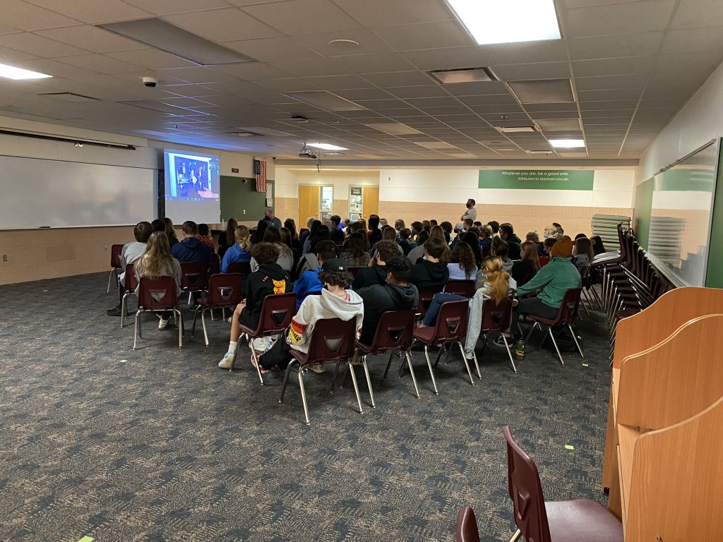
Students learning how to apply science in the real world. Credit: Charles Bittle
Investigating Bacteriophages in their Microbiome
Currently, Dr Parent is researching how viruses like bacteriophages infect hosts via host recognition and transfer of their genetic material across their cell membranes. Often, viruses recognise a specific receptor on their host that facilitates infection and so, if this receptor is altered or deleted, they are better protected. However, Dr Parent found that the S. flexneri bacteriophage, Sf6 can recognise multiple receptors and therefore, overcome Shigella defence mechanisms.
She is utilising this knowledge to investigate how the structure of Sf6 allows it to do this but also expanding her research into more complex environments. The relationship of bacteriophages with bacteria means that they can drive the evolution of their microbiome, as the bacteria try to adapt to evade the destructive effects of the tiny viruses. Dr Parent is studying the mechanisms of this phage-mediated evolution within complex microbial networks, which has scarcely been attempted before.
Summing up her fascinating research, Dr Parent says that, ‘we have made a ground-breaking discovery that Shigella phages are highly abundant in the environment. In addition, we have found some highly novel isolates that have different genome sizes, shapes, and structures compared to other previously identified bacteriophages that infect bacteria such as Salmonella and E. coli.’ Her work has opened exciting new avenues to explore in the world of bacteriophages and antibiotic resistance and will be incredibly useful for more research to come, as well as capturing the minds of budding scientists at Lincoln Southwest High School.
SHARE
DOWNLOAD E-BOOK
WATCH THE ANIMATION
REFERENCE
https://doi.org/10.33548/SCIENTIA778
MEET THE RESEARCHER
Dr Kristin N. Parent
Department of Biochemistry and Molecular Biology
Michigan State University
East Lansing, MI
USA
Dr Kristin Parent received her BSc in Molecular and Cell Biology from the University of Connecticut where she also went on to complete a PhD in Biochemistry. She conducted postdoctoral work at the University of San Diego and then took up the position of Assistant Professor at Michigan State University, where she is now Associate Professor. In addition to these achievements, Dr Parent independently established and is the Director of the RTSF Cryo-EM facility at the university and carries out her research here. Her work focuses on discovering bacteriophages in the natural environment and studying the underlying mechanisms of viral infection via host recognition and transfer of genetic material across cell membranes.
CONTACT
W: https://kparentlab.natsci.msu.edu/
Twitter: @phage4Lyfe
KEY COLLABORATORS
Instructors at Lincoln Southwest High School:
- Kevin Schrad
- Charles Bittle
- Peter Stone
Students/trainees involved with the project:
- Dr Jason Schrad
- Dr Sarah Doore
- Dr Sundharraman Subramanian
- Kendal Tinney
- Hailee Perrett
FUNDING
National Science Foundation CAREER Award (1750125)
FURTHER READING
SM Doore, JR Schrad, HR Perrett, et al., A cornucopia of Shigella phages from the Cornhusker State, Virology, 2019, 538, 45–52. DOI: https://doi.org/10.1016/j.virol.2019.09.007
SM Doore JR Schrad, WF Dean, et al., Shigella Phages Isolated during a Dysentery Outbreak Reveal Uncommon Structures and Broad Species Diversity, Journal of Virology, 2018, 92(8), e02117-17. DOI: https://doi.org/10.1128/JVI.02117-17

REPUBLISH OUR ARTICLES
We encourage all formats of sharing and republishing of our articles. Whether you want to host on your website, publication or blog, we welcome this. Find out more
Creative Commons Licence (CC BY 4.0)
This work is licensed under a Creative Commons Attribution 4.0 International License. 
What does this mean?
Share: You can copy and redistribute the material in any medium or format
Adapt: You can change, and build upon the material for any purpose, even commercially.
Credit: You must give appropriate credit, provide a link to the license, and indicate if changes were made.
SUBSCRIBE NOW
Follow Us
MORE ARTICLES YOU MAY LIKE

Elevating Histology: Rethinking Clinical Laboratory Regulations for Modern Diagnostic Demands
Histology is the science dealing with the structure and analysis of cells and their formation into tissues and organs. The profession is responsible for the preparation of all pathological tissue samples removed and collected from the human body for the microscopic detection of tissue abnormalities for disease diagnosis and treatment. Despite advances in immunohistochemistry, molecular diagnostics, and digital pathology, the US Clinical Laboratory Improvements Amendments (CLIA) regulations from 1988 have not evolved to reflect histology’s scientific demands, including performing complex diagnostic tasks essential to patient care. Elizabeth Chlipala from Premier Laboratory (LLC), Longmont, Colorado and colleagues argue for a national certification requirement and regulatory oversight for histologists. Citing current problems including quality issues, workforce shortages, and the need for standardized practices, these experts argue that recognizing histologists under CLIA would elevate the profession, ensure competency, and improve patient outcomes, challenging the current position of the College of American Pathologists.

Dr Masumi Kamiyama | Using Natural Compounds from Soy to Protect Kidneys from Damage in Diabetes
Dr Masumi Kamiyama, Associate Professor at Jumonji University, Saitama, Japan, leads research into the early detection and prevention of diabetic nephropathy, which is kidney damage caused from long-term high blood sugar that makes it harder for the kidneys to filter waste, and can potentially lead to kidney failure. Dr Kamiyama and colleagues are exploring the role of a protein called angiotensinogen (AGT) as an early biomarker of kidney damage. The team studies the potential of plant-based antioxidants, called isoflavones, to slow or halt kidney damage from highly reactive molecules, such as oxygen free radicals. Ongoing research by Professor Han Lamers (University of Oslo) and Professor Bettina Reitz-Joosse (University of Groningen) reveals how Fascist Italy weaponized ancient Rome’s language to legitimise its power and connect Mussolini’s regime to Italy’s imperial past. Their projects involve collaboration with an international team of mostly junior researchers based in Norway, the Netherlands, Austria, and Italy.

Dr JoLee Sasakamoose – Dr Mamata Pandey | Empowering Indigenous Health: The Indigenous Wellness Research Collaborative in Saskatchewan
The Indigenous Wellness Research Collaborative is a transformative alliance dedicated to advancing health systems and service delivery for Indigenous communities across Saskatchewan. Founded a decade ago and co-led by Dr Mamata Pandey, a research scientist at the Saskatchewan Health Authority, and Dr JoLee Sasakamoose (M’Chigeeng First Nation), Canadian Institute of Health Research Chair in Indigenous Wellness and Health Equity at the University of Regina, their team’s work is rooted in a commitment to Indigenous leadership and community-defined wellness goals. Guided by the Cultural Responsiveness Framework, the Collaborative prioritises creating ethical spaces that serve as a middle ground for respect, reciprocity, and authentic partnerships. The team employs a strengths-based approach to health research, centering Indigenous methodologies that respect the interconnectedness of spiritual, mental, emotional, and physical well-being.

Professor Jaya Krishnan | Revolutionary Gene Therapy Helps Hearts Regenerate After Heart Attacks
Myocardial infarction, commonly termed as a heart attack, is a major cause of death and poor health worldwide. Regenerating heart tissue is an exciting and promising concept that can have significant benefits in myocardial infarctions and related diseases, but this has not yet been achieved in real-life clinical treatments. In a collaboration between Goethe University Frankfurt and Goethe University Hospital, Professor Jaya Krishnan and colleagues address this by controlling pathologic genes involved in the development of heart failure that develops after heart attacks. The researchers demonstrate a new way of treating heart disease by aiding in the division and regrowth of heart cells after a heart attack.