
Dr Michael Hicks | Creating Skeletal Muscle from Stem Cells
Recognising the precise steps involved in the differentiation process of stem cells into various cell types is crucial to regenerative medicine. Researchers face the specific challenge of how to fine-tune and optimise protocols for directing stem cells to differentiate into specific cell lineages. As such, the directed differentiation to skeletal muscles has not yet advanced to the clinical trials stage. Dr. Michael Hicks from the School of Medicine at the University of California, Irvine, is working to change this with his extensive studies into the transition of stem cells into skeletal muscles.
Transplanting Stem Cells into Skeletal Muscles
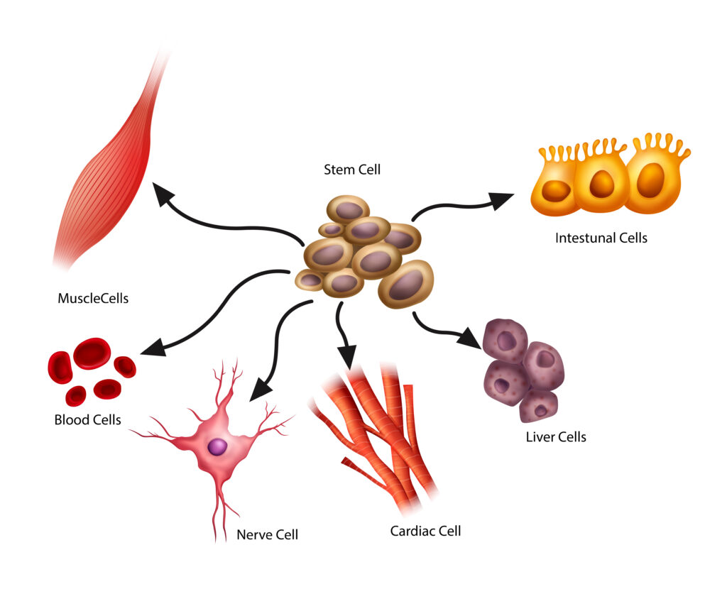
Human pluripotent stem cells (hPSCs) have a remarkable ability to differentiate into any cell type in the body, including muscle cells, blood cells, and neurons. Unlike human embryonic stem cells, induced hiPSCs are derived from adults, often from individuals with specific genetic disorders, and offer a promising avenue for tailoring personalised regenerative medicine strategies for a multitude of diseases. For instance, the directed differentiation of hPSCs into pancreatic cells producing insulin has revolutionised the treatment landscape for Type 1 diabetes. Furthermore, hPSC-based therapies hold immense potential for addressing a surprising spectrum of debilitating conditions, ranging from neurodegenerative disorders like Parkinson’s and Alzheimer’s diseases to genetic ailments, as well as diverse diseases like arthritis and cardiac defects.
The process of transplanting stem cells into skeletal muscles is of particular interest in medical science. The ability of stem cells to self-renew could offer an alternative response to injuries and replace damaged muscles over a lifetime. However, so far, generating clinical-grade skeletal muscle cells from hPSCs has been challenging.
Obstacles and Gaps
Researchers have made progress in isolating hPSCs from patients and modifying them in the laboratory. However, challenges arise when reintroducing these modified cells into patients, where they must effectively replicate and regenerate. Dr Michael Hicks from the School of Medicine at the University of California, Irvine, is working to minimze the gaps in understanding the progenitors involved in early skeletal muscle formation, especially during initial myogenic commitment.
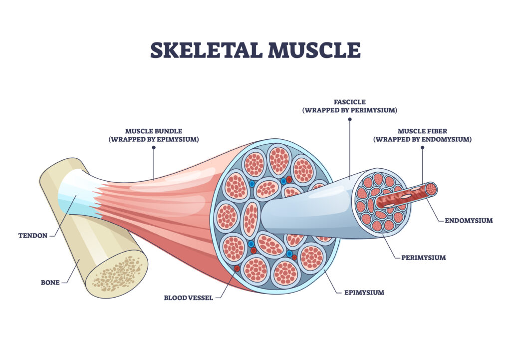
To form the skeletal muscle system, progenitors first divide to form paraxial mesoderm, which develops into blocks of tissue known as somites. These blocks of tissue then divide into the ventral sclerotome giving rise to bone and cartilage, and the dorsal dermomyotome from which skeletal muscles arise. However, genetic variability, that is, differences in the sequences of genes between individuals, adds complications when attempting to do this in a dish since small differences between cells taken from different people can lead to inconsistent outcomes during this differentiation process.
Crucial transcription factors (proteins involved in converting or transcribing DNA into RNA that composes our genes) like Pax3, Pax7, Eya1, and Six1 play are involved in the process of muscle development. Still, their role and timing in hPSC differentiation remains unclear. Additional signaling mechanisms like the Wnt pathway and other secretions from neighboring cells also play a part in directing muscle cell fates.
Dr Hicks emphasises that research development should focus on understanding in vitro myogenesis which focuses on the robust differentiation of hPSCs into pre-myogenic progenitors to identify optimal microenvironments for muscle development.
Identifying Early Differentiation Checkpoints
Dr Hicks recognised that studying prenatal muscle development might help address the gaps in understanding hPSC muscle differentiation. His team examined progenitor markers expressed in the developing limb at the seventh week of human embryos, leading to the identification of a SIX1+PAX3+ co-expressing myogenic population. Further investigation revealed that SIX1 expression could serve as a potential checkpoint to distinguish between hPSC differentiations that would later successfully mature to skeletal muscle progenitor cells.
The expression levels of SIX1 can be used to optimise protocols for directed differentiation of myogenic cells derived from hPSCs in clinical settings. By inducing robust expression of SIX1+PAX3+ precursors early in directed differentiations, the authors reported consistent derivation of muscle formation across all patient hiPSC lines tested. In the absence of SIX1, PAX3 expression was affected in cell lines, and muscle progenitors failed to emerge. The researchers propose predicting the emergence of the SIX1+PAX3+ phenotype as an early indicator for improving subsequent myogenic differentiation.
Building on Ground-breaking Discoveries
The ability to induce hPSCs derived from any individual and to reprogramme them to an embryonic state in laboratory settings was a ground-breaking discovery by Drs. Yamanaka and Gurdon that won the Nobel Prize in 2007. These reprogrammed cells hold the potential to be differentiated into various tissue types, offering immense potential for personalised medicine.
Achieving consistent differentiation of induced hPSCs is crucial for generating clinical-grade muscle cells, which are essential for effective cell therapy to replace damaged or dysfunction muscle tissues. Dr Hicks’ latest study presents a stepwise approach to optimise differentiation protocols for deriving skeletal muscle progenitor cells from various patient specific hPSCs lines. The team identified a critical developmental checkpoint at an early stage of myogenic commitment that predicts successful differentiation and validated the emergence of myogenic-specific markers early in the protocol.
The next step in Dr Hick’s research is to test and develop strategies to mature skeletal muscle progenitor cells into satellite cells like those found in adults. The idea is that these satellite cells can reside in the body’s stem-cell niche and continuously self-renew damaged muscles. Such regenerative cell replacement therapy holds significant promise for personalised medicine across multiple medical specialities.
SHARE
DOWNLOAD E-BOOK
REFERENCE
https://doi.org/10.33548/SCIENTIA1160
MEET THE RESEARCHER

Dr Michael Hicks
Department of Physiology and Biophysics
School of Medicine
University of California, Irvine
Irvine, CA
USA
Dr Michael Hicks is an Assistant Professor of Physiology and Biophysics at the University of California, Irvine (UCI). He also serves as associate director of the UCI Muscle Biology and Disease Research Center and contributes to the Sue and Bill Gross Stem Cell Research Center. His research focuses on skeletal muscle regeneration using adult muscle stem cells and human pluripotent stem cells. He completed his doctoral research at the University of Arizona, then undertook research at the University College London and the University of California, Los Angeles. His research group studies stem cell regeneration and the influence of diseased microenvironments on stem cell behaviour by employing emerging technologies. Dr Hicks holds two patents and has several notable publications, including papers in Stem Cell Stem and Nature Cell Biology.
CONTACT
E: mrhicks1@hs.uci.edu
W: https://hickslab.org/
X: https://twitter.com/mic_hicks/
KEY COLLABORATORS
Olga Jaime, School of Medicine, University of California, Irvine
Jessica Arias, School of Medicine, University of California, Irvine
Dr Shreya Pavani, School of Medicine, University of California, Irvine
Professor April Pyle, Microbiology, Immunology, and Molecular Genetics, University of California, Los Angeles
Please add any additional collaborators you wish to acknowledge here (name and institute)
FUNDING
National Institutes of Health
FURTHER READING
OG Jaime, J Arias, S Pavani, et al., SIX1+PAX3+ identify a progenitor for myogenic lineage commitment from hPSCs, Development, 2023, 150(14), dev201509. DOI: https://doi.org/10.1242/dev.201509
REPUBLISH OUR ARTICLES
We encourage all formats of sharing and republishing of our articles. Whether you want to host on your website, publication or blog, we welcome this. Find out more
Creative Commons Licence (CC BY 4.0)
This work is licensed under a Creative Commons Attribution 4.0 International License. 
What does this mean?
Share: You can copy and redistribute the material in any medium or format
Adapt: You can change, and build upon the material for any purpose, even commercially.
Credit: You must give appropriate credit, provide a link to the license, and indicate if changes were made.
SUBSCRIBE NOW
Follow Us
MORE ARTICLES YOU MAY LIKE

Assoc Prof. Nicholas Brown | Rethinking Prostate Care: A New Frontier in Treating Benign Prostatic Hyperplasia
For millions of men, ageing brings with it a set of frustrating and often disruptive urinary symptoms. These symptoms, caused by benign prostatic hyperplasia, or BPH, can affect sleep, confidence, and overall quality of life. Traditionally, treatment follows a familiar path. Patients begin with medications, often for years, and may eventually progress to surgery if symptoms worsen. Yet this pathway is not without its drawbacks. Medications can cause side effects, while surgery carries risks and recovery time. In recent years, a minimally invasive interventional radiology procedure called prostate artery embolisation, or PAE, has begun to challenge this traditional model. At the forefront of this shift is a collaborative research group, led by Dr. Nicholas Brown of the University of Queensland, whose series of P-EASY studies has explored whether PAE could transform how BPH is treated, particularly at earlier stages.

Jean Lycke | Addressing Unmet Medical Needs in Mucosal Disease: A Close-to-Market Innovation Approach
Recurrent Aphthous Stomatitis (RAS) is an oral condition characterized by one or several painful mucosal ulcers. RAS affects a large proportion of the population and has a point prevalence of approximately 2–3%, daily. The etiology remains unknown, and there is currently no curative treatment. Most patients experience recurring episodes over time, with each episode typically lasting up to a week. Here, we describe the development of a mucoadhesive patch which, when applied over a RAS ulcer, provides rapid pain relief. The patch is easy for patients to apply when symptoms begin and has the potential to be used as an over-the-counter product. The development of the Mucocort mucoadhesive patch is an example of a Close-to-Market innovation strategy that embraces simplicity within a complex healthcare system. By simplifying the product concept, the team has reduced the number of regulatory steps required before market approval. This MedTech/Pharma innovation model, known as the “4R” framework – Re-purposing, Re-formulation, Re-positioning, and Re-patenting – has guided the program from concept to commercialization. In addition to the biodegradable mucoadhesive patch developed for RAS ulcers, the team is extending the innovation concept to a mucoadhesive gel formulation for the prevention and treatment of chemotherapy-induced mucositis. This gel-based program is being commercialized separately through MucoShield.
The Translational Asian Agerelated Macular Degeneration Program Phase 2 (TAAP-2): Reimagining the Future of Vision Care
Age-related macular degeneration, often abbreviated as AMD, is one of the leading causes of vision loss among older adults worldwide. In Asia, where populations are ageing rapidly, its impact is particularly profound. For many, the disease quietly erodes central vision, making everyday activities such as reading, driving, and recognising faces increasingly difficult. Against this backdrop, the Translational Asian Age-related Macular Degeneration Programme, or TAAP for short, has emerged as a bold and ambitious effort to confront the disease headon. Now in its second phase, TAAP-2 represents a significant evolution in both scientific scope and clinical ambition.

Ms. Aikaterini Dritsoula | Looking Beyond Snoring: How Hidden Airway Problems Shape Children’s Sleep
For many parents, a child’s snoring may seem harmless, even endearing. Yet in some cases, it signals something more serious. Obstructive sleep apnoea is a condition in which a child’s breathing is repeatedly disrupted during sleep. These interruptions can affect growth, behaviour, and learning. Children with this condition may toss and turn at night, struggle to concentrate during the day, or show signs of hyperactivity and fatigue. Traditionally, enlarged tonsils and adenoids have been seen as the main culprits. Surgery to remove them has long been considered the standard treatment. However, research led by Consultant ENT Surgeon Ms. Aikaterini Dritsoula of The Leeds Teaching Hospitals NHS Trust invites us to look deeper. Her work suggests that the story is often more complex, especially in very young children.