
Dr. Petr Kelbich | Investigating Inflammatory Conditions in Extravascular Body Fluids: An Important New Parameter
Diagnosing disorders of the brain and other organs can often feel like solving a challenging puzzle. Analyzing non-blood body fluids provides valuable clues that can help address this complexity. To enhance this process, Dr. Petr Kelbich from Jan Evangelista Purkyně University and Masaryk Hospital in Ústí nad Labem, Czech Republic, introduced an innovative method called Cytological-Energy Analysis.
At the core of this approach is the Coefficient of Energy Balance (KEB), a mathematical concept that offers deeper insights into immune cell activity and energy requirements during inflammation. By using this method, doctors can identify issues in different organs with greater precision, making diagnoses more accurate and efficient.
Dr. Kelbich initially introduced this method for diagnosing central nervous system (CNS) disorders through cerebrospinal fluid (CSF) analysis.
Cytological Investigation of CSF and the Coefficient of Energy Balance (KEB)
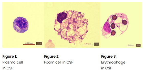
The first step of this method involves a cytological analysis of CSF. The cells found in CSF can offer valuable insights and help detect various CNS disorders. For example:
- Plasma cells (Figure 1): Associated with antibody production during inflammation.
- Foam cells (Figure 2): Indicating tissue damage.
- Erythrophages (Figure 3): Suggesting bleeding in the CSF compartment.
- Tumor cells (Figure 4): Pointing to malignant infiltration of the meninges.
- Different pathogens (Figures 5 and 6): Pointing to infection in CNS.

However, simply observing the cells isn’t always enough. That’s where the Coefficient of Energy Balance (KEB) plays a crucial role—it calculates energy production in the affected area by analyzing glucose and lactate molar concentrations. This reveals the activity of immune cells and the severity of inflammation.
The KEB formula:

This formula enhances these insights by estimating the production of adenosine triphosphate (ATP), offering a clearer understanding of metabolic activity and energy demands within the CSF (Figure 7).
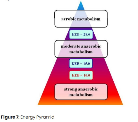
The energy pyramid illustrates the energy dynamics in normal and inflamed CSF conditions.
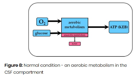
Under normal conditions, CSF metabolism is predominantly aerobic, yielding high ATP production and KEB values above 28.0 (Figure 8).
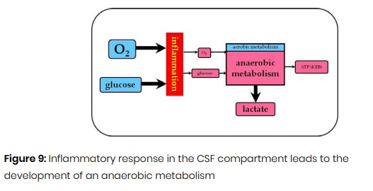
During inflammation, immune cell activation increases glucose and oxygen consumption, shifting to anaerobic metabolism. This change can result in reduced ATP production and lower KEB values (Figure 9):
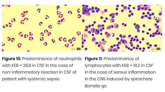
- KEB values above 28.0 indicate aerobic metabolism, which is typical in the absence of inflammation or during mild serous inflammation (Figure 10).
- KEB values between 28.0 and 15.0 suggest moderate anaerobic metabolism, commonly reflecting the increased energy demands of an activated immune system during a “serous” inflammatory response (Figure 11).
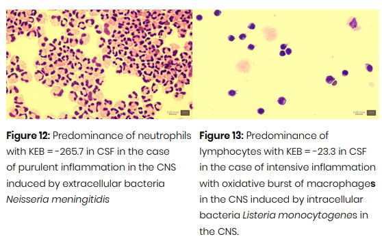
- KEB values below 10.0 signify strong anaerobic metabolism, typically associated with an intense inflammatory response. This process is characterized by the rapid release of reactive oxygen species by immune cells to combat pathogens, a phenomenon known as the oxidative burst.
The oxidative burst of neutrophils is central to purulent inflammation, a reaction typically triggered by extracellular bacteria in the CNS (Figure 12). In contrast, the oxidative burst of macrophages is crucial for combating intracellular pathogens, fungal infections, or tumor development (Figure 13).
Extending Cytological-Energy Applications
By combining cytological and energy analyses, the KEB method offers a more precise understanding of inflammation across various organ systems (Figures 14–16).
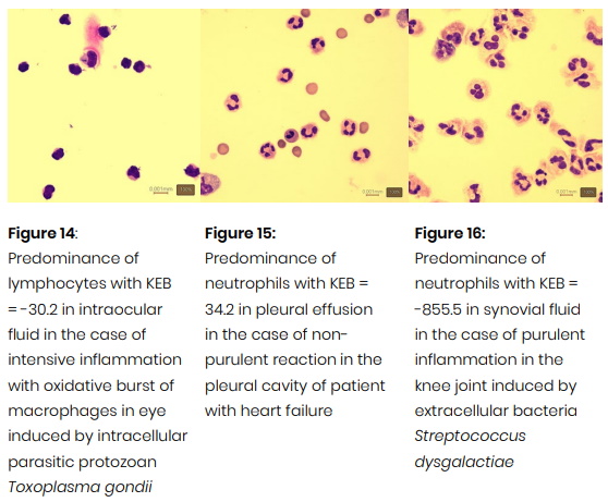
Following successful application in cerebrospinal fluid (CSF) analysis, Dr. Kelbich expanded his collaboration with medical specialists to explore other extravascular body fluids. These included pleural, pericardial, and abdominal effusions, as well as intraocular fluid, peritoneal dialysate, amniotic fluid, synovial fluid, and others. The results were highly promising, demonstrating that cytological-energy analysis has significant potential for broader applications across multiple medical fields.
Towards More Precise Diagnostics
Dr. Kelbich’s approach integrates cytological and metabolic profiling, providing a comprehensive framework for diagnosing inflammatory conditions across various organ systems. By quantifying the type and intensity of immune responses, Cytological-Energy Analysis enables:
- More precise differentiation between types of local inflammation.
- Enhanced monitoring of disease progression.
- More targeted treatment strategies, leading to improved patient outcomes.
This method seamlessly combines scientific knowledge with practical application, offering deeper insights into localized bodily processes. It represents a promising tool to advance the diagnosis and treatment of a wide range of conditions, ultimately benefiting patients worldwide.
SHARE
DOWNLOAD E-BOOK

REFERENCE
https://doi.org/10.33548/SCIENTIA1243
MEET THE RESEARCHER

Dr. Petr Kelbich
Jan Evangelista Purkyně University and Masaryk Hospital in Ústí nad Labem, Czech Republic
Dr. Petr Kelbich serves as the head of the Department of Biomedicine and Laboratory Diagnostics at Jan Evangelista Purkyně University and Masaryk Hospital in Ústí nad Labem, Czech Republic. His primary research focuses on the analysis of cerebrospinal fluid and other extravascular body fluids to enhance the diagnosis of central nervous system disorders and diseases affecting other organ systems. He developed an accessible diagnostic procedure aimed at refining diagnostic accuracy across various medical fields. Dr. Kelbich presented his work to the global scientific community about a decade ago and has since contributed to the field through peer-reviewed publications and conference presentations, sharing advancements in diagnostic methodologies.
CONTACT
E: petr.kelbich@kzcr.eu
W: https://fzs.ujep.cz/cs/ustav-biomediciny-a-laboratorni-diagnostiky
FUNDING
Internal Grants of the Krajská zdravotní, a.s. in Ústí nad Labem, Czech Republic: IGA-KZ-2021-1-1, IGA-KZ-2021-1-2, IGA-KZ-2020-1-7, IGA-KZ-2019-1-7, IGA-KZ-2019-1-9, IGA-KZ-2017-1-5, and KZ-2016-1-9.
FURTHER READING
P Kelbich, et al., The Cytological Energy Detection of Purulent Inflammation in Synovial Fluid Is Not All Black and White, Biomedicines, 2024, 12(3), 667. DOI: https://doi.org/10.3390/biomedicines12030667
P Kelbich, et al., Basic Analysis of the Cerebrospinal Fluid: An Important Framework for Laboratory Diagnostics of the Impairment of the Central Nervous System, Current Issues in Molecular Biology, 2022, 44, 3666–3680. DOI: https://doi.org/10.3390/cimb44080251
P Kelbich, et al., Neutrophils in Extravascular Body Fluids: Cytological-Energy Analysis Enables Rapid, Reliable and Inexpensive Detection of Purulent Inflammation and Tissue Damage, Life, 2022, 12, 160. DOI: https://doi.org/10.3390/life12020160
E Vanaskova, et al., Reactive synovitis of the knee joint after COVID-19 vaccination: The first ultrastructural analysis of synovial fluid, International Journal of Rheumatic Diseases, 2022, 00, 1-4. DOI: https://doi.org/10.1111/1756-185X.14411
E Vanaskova, et al., Malignant Knee Joint Effusion – A New Dimension of Laboratory Diagnostics, Applied Sciences, 2022, 12, 994. DOI: https://doi.org/10.3390/app12030994
P Kelbich, et al., Development of the Cerebrospinal Fluid in Early Stage after Hemorrhage in the Central Nervous System, Life, 2021, 11, 300. DOI: https://doi.org/10.3390/life11040300
J Soukup, et al., Toxocariasis as a Rare Parasitic Complication of a Transthoracic Spine Surgery Procedure, Medicia, 2021, 57, 1328. DOI: https://doi.org/10.3390/medicina57121328
P Kelbich, et al., Can Aspartate Aminotransferase in the Cerebrospinal Fluid be a Reliable Predictive Parameter?, Brain Sciences, 2020, 10, 698. DOI: https://doi.org/10.3390/brainsci10100698
I Matuchova, et al., Cytological-energy analysis of pleural effusion with predominance of neutrophils, Therapeutic Advances in Respiratory Disease, 2020, 14, 1–10. DOI: https://doi.org/10.1177/1753466620935772
P Kelbich, et al., Cytological-energy analysis of pleural effusions, Annals of Clinical Biochemistry, 2019, 56(6), 630–637. DOI: https://doi.org/10.1177/0004563219845415
P Kelbich, et al., Principles of the Cytological-Energy Analysis of the Extravascular Body Fluids, Biochemistry & Molecular Biology Journal, 2017, 3(1), 6. DOI: 10.21767/2471-8084.100031
P Kelbich, et al., Coefficient of energy balance, a new parameter for basic investigation of the cerebrospinal fluid, Clinical Chemistry and Laboratory Medicine, 2014, 52(7), 1009–1017. DOI: https://doi.org/10.1515/cclm-2013-0953
![]()
REPUBLISH OUR ARTICLES
We encourage all formats of sharing and republishing of our articles. Whether you want to host on your website, publication or blog, we welcome this. Find out more
Creative Commons Licence (CC BY 4.0)
This work is licensed under a Creative Commons Attribution 4.0 International License. 
What does this mean?
Share: You can copy and redistribute the material in any medium or format
Adapt: You can change, and build upon the material for any purpose, even commercially.
Credit: You must give appropriate credit, provide a link to the license, and indicate if changes were made.
SUBSCRIBE NOW
Follow Us
MORE ARTICLES YOU MAY LIKE

Jean Lycke | Addressing Unmet Medical Needs in Mucosal Disease: A Close-to-Market Innovation Approach
Recurrent Aphthous Stomatitis (RAS) is an oral condition characterized by one or several painful mucosal ulcers. RAS affects a large proportion of the population and has a point prevalence of approximately 2–3%, daily. The etiology remains unknown, and there is currently no curative treatment. Most patients experience recurring episodes over time, with each episode typically lasting up to a week. Here, we describe the development of a mucoadhesive patch which, when applied over a RAS ulcer, provides rapid pain relief. The patch is easy for patients to apply when symptoms begin and has the potential to be used as an over-the-counter product. The development of the Mucocort mucoadhesive patch is an example of a Close-to-Market innovation strategy that embraces simplicity within a complex healthcare system. By simplifying the product concept, the team has reduced the number of regulatory steps required before market approval. This MedTech/Pharma innovation model, known as the “4R” framework – Re-purposing, Re-formulation, Re-positioning, and Re-patenting – has guided the program from concept to commercialization. In addition to the biodegradable mucoadhesive patch developed for RAS ulcers, the team is extending the innovation concept to a mucoadhesive gel formulation for the prevention and treatment of chemotherapy-induced mucositis. This gel-based program is being commercialized separately through MucoShield.
The Translational Asian Agerelated Macular Degeneration Program Phase 2 (TAAP-2): Reimagining the Future of Vision Care
Age-related macular degeneration, often abbreviated as AMD, is one of the leading causes of vision loss among older adults worldwide. In Asia, where populations are ageing rapidly, its impact is particularly profound. For many, the disease quietly erodes central vision, making everyday activities such as reading, driving, and recognising faces increasingly difficult. Against this backdrop, the Translational Asian Age-related Macular Degeneration Programme, or TAAP for short, has emerged as a bold and ambitious effort to confront the disease headon. Now in its second phase, TAAP-2 represents a significant evolution in both scientific scope and clinical ambition.

Ms. Aikaterini Dritsoula | Looking Beyond Snoring: How Hidden Airway Problems Shape Children’s Sleep
For many parents, a child’s snoring may seem harmless, even endearing. Yet in some cases, it signals something more serious. Obstructive sleep apnoea is a condition in which a child’s breathing is repeatedly disrupted during sleep. These interruptions can affect growth, behaviour, and learning. Children with this condition may toss and turn at night, struggle to concentrate during the day, or show signs of hyperactivity and fatigue. Traditionally, enlarged tonsils and adenoids have been seen as the main culprits. Surgery to remove them has long been considered the standard treatment. However, research led by Consultant ENT Surgeon Ms. Aikaterini Dritsoula of The Leeds Teaching Hospitals NHS Trust invites us to look deeper. Her work suggests that the story is often more complex, especially in very young children.

Professor Neil Coffee – Professor Vincent Versace | Mapping Health Access: Using Address-Level Intelligence for Smarter Services
Accessing healthcare is a serious challenge for people living in rural and remote Australia. Large distances, sparse populations, and limited services can prevent residents from receiving care when they need it. Professors Neil Coffee and Vincent Versace at Deakin University’s Centre for Australian Research into Access (CARA) are leading research to model healthcare service access across the country, to provide new insights that can guide health planning and policy, as well as other services such as education. This work combines the curation of detailed address level residential dwellings and road network data to calculate access to service metrics (time and distance). These metrics are applied to the simulated residential dwelling population, to quantify the population with poor access to health services.