
Improving Joint Replacement Materials
Hip and knee replacements are key for the health and well-being of many people. Unfortunately, the materials used in these procedures are known to wear and corrode once placed within the human body. Dr Stefano Mischler and his team from École Polytechnique Fédérale de Lausanne, together with Professor Dr Brigitte Jolles-Haeberli and her team from Lausanne University Hospital in Switzerland, have conducted exciting new research that advances our understanding of the factors influencing corrosion in the human body. Their work has exciting implications for the future monitoring of implant conditions, particularly valuable in the face of our increasingly ageing population.
Joint Replacement Surgery
Joint replacement surgeries are among the most common elective medical interventions in people over 60 years old. They are performed to help people regain the use of joints, including hips and knees, which have been damaged by degenerative diseases such as osteoarthritis or trauma. Over a million people in the USA undergo hip or knee replacement surgery each year, and with global populations increasingly ageing and increasing high-level recreational sports, this type of surgery is going to become more common around the world.
In this type of surgery, the damaged parts of the joint are replaced with high-quality man-made materials such as metal alloys, including titanium and cobalt-chromium-molybdenum (CoCrMo), primarily used for their strength and resilience.
Joint replacement surgery helps improve people’s quality of life by reducing pain, restoring mobility and, subsequently, their independence. However, the materials used do not last forever in the body, and the average hip replacement lasts approximately 20 years before additional surgery is needed to either repair or replace it.
To improve joint replacement surgery, Dr Stefano Mischler and a team of scientists from École Polytechnique Fédérale de Lausanne and Professor Dr Brigitte Jolles-Haeberli at Lausanne University Hospital set up an important collaboration. Their goal is to revolutionalise understanding of how these materials corrode within the body and to develop materials that last as long as possible, thus reducing the need for additional surgery in people with joint replacements.
Implant Corrosion in the Body
To improve the performance and longevity of these implant materials, it is essential to understand how they deteriorate in the human body. So far, studies investigating these metals have only been performed in laboratory studies using simulated body fluids – which may not accurately represent the conditions in the body. These laboratory studies have shown that the durability of these metals is dependent on a wide range of factors (including those relating to the individual patient). Furthermore, the different factors have only been studied individually. As such, the interactions between various factors as they may occur in the body are not yet fully understood.
To address this knowledge gap, the collaborative team of scientists and clinicians conducted a series of experiments to investigate the corrosion behaviour of implant materials in real human conditions to help predict the lifetime of joint replacement implants.
Professor Dr Jolles-Haeberli, a skilled hip and knee surgeon, extracted synovial fluid (the liquid found in joints that helps them move smoothly) from more than 170 patients during planned elective arthroplasty with patient consent. The pH, colour, and viscosity of the extracted fluid were assessed to help understand how the materials react in different patients. The ability of the synovial fluid to conduct an electrical charge allowed the use of electrochemical techniques to investigate the interaction between the implant materials and the synovial fluid and determine how likely the materials were to corrode and how fast this would happen. The obtained corrosion rates from experiments (proportional to the metal ion release from the implant material into the human body) were compared to the metal ion levels in patients’ urine and found to correspond very well. Therefore, these new experiments could be considered a good marker for the reactivity of a patient toward specific implant material.
Titanium and CoCrMo are often combined in implants. For example, a hip replacement may be made up of a CoCrMo ball attached to a titanium stem. In theory, this combination may increase corrosion in the body if one material is more noble than the other – otherwise known as galvanic corrosion. With the present data, no significant risk of galvanic corrosion in synovial fluid was observed between CoCrMo and Titanium.
Alongside electrochemical corrosion, joint implants are also affected by physical friction and wear and tear caused by movement and mechanical loading. The mechanical wear damages the metal implant, allowing the potentially corrosive synovial fluid to enhance its effect and increase the implant damage rate. The combined effect is known as tribocorrosion and is one of the main causes of joint implant degradation. Dr Mischler is an expert in this phenomenon, and the team combined tribocorrosion models with their electrochemical experiments in synovial fluid to calculate the overall degradation rates of hip replacements.

Implant Material Corrosion Rates are Patient-specific
A key finding (supporting the team’s previous work) was that corrosion rates of both titanium and CoCrMo implant material vary with the patient in which it is implanted. The team found that the patient’s synovial fluid impacts the lifespan of metals used as implants in multiple ways. Some molecules in the fluid react with the implant and increase the degradation rate, whereas other molecules, such as proteins, can stick to the implant, protecting it from corrosion. They found no risk of galvanic corrosion between the two types of metal as they have very similar electrical properties when submerged in human synovial fluid.
The team also reported that corrosion rates in the study correlated well with levels of metal ions in the patient’s urine. This is a particularly exciting finding as it may be used in the future to understand how long an implant may last in an individual patient, reducing the need for more invasive techniques.
Electrochemical Techniques in Clinical Medicine
The unique collaboration between École Polytechnique Fédérale de Lausanne and Lausanne University Hospital has highlighted the value of electrochemical testing in investigating the longevity of joint replacement implant material. The team’s findings show that metal implant degradation rates are specific to individual patients due to variations in synovial fluid and physical wear factors. This means that the same implant will last for a different amount of time in different patients and emphasises the need for a non-invasive way to measure implant condition. The team’s work demonstrates that the corrosion rates of both titanium and CoCrMo implants in human synovial fluid match closely with the rate at which metal ions are released into the patients. Importantly, this suggests that testing the level of metal ions in patients’ bodies through a simple urine test could be used to monitor the condition and corrosion of their joint replacement implant.
SHARE
DOWNLOAD E-BOOK
REFERENCE
https://doi.org/10.33548/SCIENTIA1118
MEET THE RESEARCHERS
École Polytechnique Fédérale de Lausanne, Institute of Materials, Tribology and Interface Chemistry, Lausanne, Switzerland
Dr Stefano Mischler
Dr Stefano Mischler obtained his PhD from the Swiss Federal Institute of Technology, where he is now head of the Tribology and Interface Chemistry Unit, where he leads research on surface chemical effects in tribology, biotribology and biocorrosion, tribology in microfabrication processes, and wear protection methods. He has published over 100 peer-reviewed journal articles and several book chapters. In addition to his research, Dr Mischler provides PhD and MSc supervision and teaches both undergraduate and postgraduate courses.
CONTACT

Dr Anna Igual Munoz
Dr Anna Igual Munoz received her PhD from the Technical University of Valencia and has subsequently worked at the University of Virginia and École Polytechnique Fédérale de Lausanne, where she is currently a senior scientist responsible for research projects related to electrochemical corrosion and tribology. Dr Igual Munoz sits on the Editorial board of the Journal of Bio- and Tribo-Corrosion and is a reviewer of the Spanish National Foundation and the European Commission. She also supervises and teaches at undergraduate and postgraduate levels and has eight teaching-related publications.
CONTACT
E: anna.igualmunoz@epfl.ch

Dr Yueyue Bao
After obtaining an MSc from the University of Science and Technology Beijing, Dr Yueyue Bao was awarded a PhD in the electrochemical behaviour of Ti and CoCrMo alloy in human synovial fluids. Dr Bao’s main research interests are focused on metal and implant materials, tribocorrosion, and electrochemistry. She has presented her work at international conferences. She has received multiple awards and scholarships for her work, including the Miyoshi Outstanding Graduate Student from the University of Science and Technology Beijing and an Academic Merit Scholarship from Hohai University.
CONTACT
Professor Dr Brigitte Joles-Haeberli and team, Lausanne University Hospital, Switzerland
Professor Dr Brigitte Jolles-Haeberli founded the Swiss BioMotion Lab (SBML) with Dr Julien Favre in 2014, and continues to lead this laboratory with him. In parallel to her academic activities, she is a senior consultant orthopaedic surgeon in hip and knee surgery at the Hirslanden clinics in Lausanne and the COO of the Department of Musculoskeletal Medicine (DAL) at CHUV. In 2023, she was elected to the renowned French Academy of Surgery in recognition of decades of excellence in orthopaedics as a surgeon, instructor, and researcher.
CONTACT
E: brigitte.jolles-haeberli@chuv.ch
FUNDING
Founds National Suisse
FURTHER READING
Y Bao, A Igual Munoz, B Jolles, S Mischler, Assessment of in-vivo corrosion of Ti and CoCrMo joint implants by electrochemical measurements in human synovial liquids, Electrochimica Acta, 2024, 476, 143708. DOI: https://doi.org/10.1016/j.electacta.2023.143708
![]()

REPUBLISH OUR ARTICLES
We encourage all formats of sharing and republishing of our articles. Whether you want to host on your website, publication or blog, we welcome this. Find out more
Creative Commons Licence (CC BY 4.0)
This work is licensed under a Creative Commons Attribution 4.0 International License. 
What does this mean?
Share: You can copy and redistribute the material in any medium or format
Adapt: You can change, and build upon the material for any purpose, even commercially.
Credit: You must give appropriate credit, provide a link to the license, and indicate if changes were made.
SUBSCRIBE NOW
Follow Us
MORE ARTICLES YOU MAY LIKE
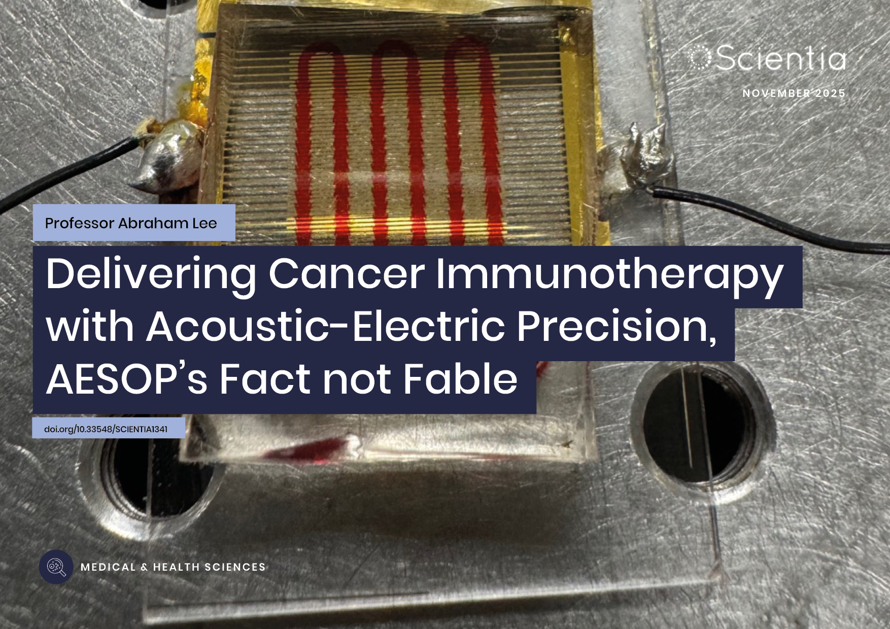
Professor Abraham P. Lee | Delivering Cancer Immunotherapy with Acoustic-Electric Precision, AESOP’s Fact not Fable
Chimeric Antigen Receptor (CAR) T-cell therapy offers life-saving potential, particularly against blood cancers, but severe side effects such as cytokine release syndrome (CRS) limit its safety. These toxicities are linked to uncontrolled CAR expression levels on the T-cell surface. Led by Professor Abraham P. Lee, researchers at the University of California, Irvine, have developed an advanced microfluidic system, called the Acoustic-Electric Shear Orbiting Poration (AESOP) platform, to precisely control the dose of genetic material delivered into primary T cells. This innovation promises safer, more homogeneous, and highly effective cellular immunotherapies.

Dr Ray Stewart | Barriers to Dental Care for People with Special Needs: A Crisis of Neglect and Inaction
For people with special healthcare needs, something as basic as visiting a dentist can be nearly impossible. A ground-breaking paper by researchers at the University of California, San Francisco (UCSF) exposes the scale of this crisis. By outlining potential paths forward, Dr Ray Stewart and Dr Ben Meisel offer hope for significant improvements in access to essential dental care.
Dr Liisa Laakso | Lighting the Way: Exploring Photobiomodulation to Ease MELAS
MELAS is a rare and serious genetic condition that affects how the body’s cells produce energy, leading to extreme fatigue, muscle weakness, and a range of other symptoms. With no cure currently available, treatment focuses only on managing complications.
A team of researchers led by Dr Liisa Laakso at the Mater Research Institute-University of Queensland, Australia, is exploring an innovative, non-drug therapy called photobiomodulation, which uses light to stimulate mitochondria to work more efficiently. This pioneering study will provide intial evidence on whether PBM can safely reduce fatigue and improve quality of life for people living with MELAS, paving the way for future clinical trials.

Professor John Paul Pezacki, PhD, FRSC (UK) | Engineering Proteins for the Prevention of Disease Progression
The way in which viruses invade and replicate within their hosts involves a multilayered system of protein-based interactions, and understanding the mechanisms at play is crucial when developing potential treatments. Utilising new techniques such as genetic code expansion, Professor John Paul Pezacki and his team of researchers at the University of Ottawa in Canada have designed a novel, highly specific artificial protein complex which can halt the progression of viral infections in human cells. They have identified and described a novel approach to wider preventative and restorative therapeutics in human disease.