
Professor Nancy Burnham | Imaging on the Nanoscale: Improving Techniques in Atomic Force Microscopy
Article written by Imogen Forbes, MSci
Atomic force microscopy (AFM) provides the means to image surfaces with nanometre resolution, allowing scientists to look at the individual building blocks and forces that make up the world around us. Professor Nancy Burnham of Worcester Polytechnic Institute and her colleagues Lei Lyu and Lily Poulikakos at the Swiss Federal Laboratories for Materials Science and Technology (Empa) have worked on how we can reduce artefacts in these images and ensure they are accurately interpreted. By considering and applying these techniques, high-quality AFM research can be produced.
What is Atomic Force Microscopy?
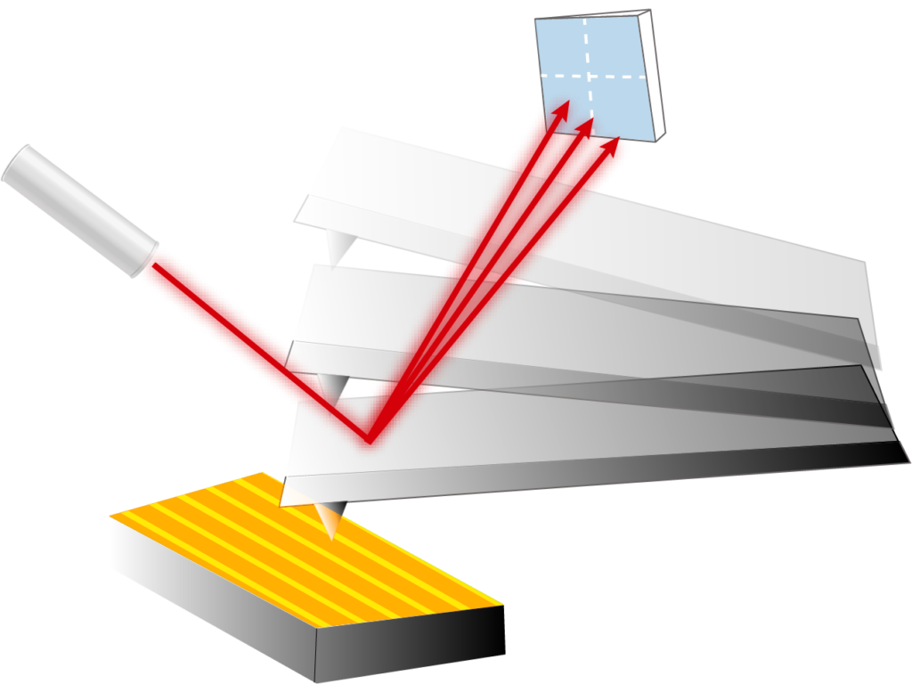
Atomic force microscopy (AFM) is used to help us learn more about a diverse range of materials, from asphalt to biological samples, through accurately imaging the surface of the material. To carry this out, AFM uses a cantilever with a sharp tip on one end. The forces from the sample affect the tip, and the cantilever bends. The bending is detected by shining a laser onto the cantilever, and measuring the light reflected from it using a photodetector. The tip is then scanned over the whole sample, allowing an image to be built from the variation in the light detected at the photodetector.
Professor Nancy Burnham of Worcester Polytechnic Institute and her colleagues highlight the importance of the correct presentation and explanation of images from AFM. These images can contain artefacts, or undesirable patterns or distortions, which can either obscure the image or contribute to the result being misinterpreted. Their work offers practical solutions to help avoid these artefacts, thus improving the outcomes from AFM imaging.
Image Processing in AFM
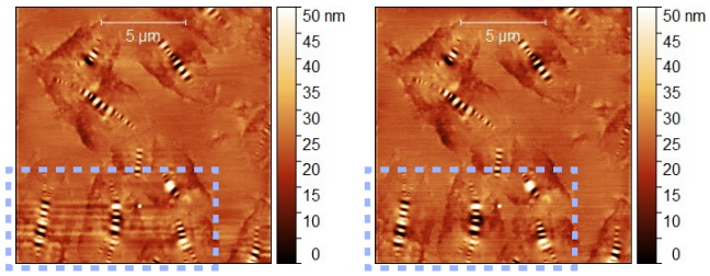
With AFM, image processing techniques are often required to help process the data, but these can sometimes result in artefacts. Professor Burnham’s team identified how distortions can occur due to a technique called row alignment. They demonstrate this by imaging ‘bee’ structures in bitumen, which is used to bind asphalt. The bee structures are seen as regions of dark and light stripes in the image of the bitumen sample.
As the tip of the AFM set-up scans over a bee structure in a row, it may see some grey pixels due to the regions of the sample without a bee, followed by some lighter pixels as a result of the light stripe of a bee. In the next row, it may see some grey pixels, followed by some darker pixels as a result of the bee’s dark stripe. In a standard row alignment process, each row’s average pixel colour is aligned with the average greyscale colour in the image. This means that the rows which contain the lighter pixels are set to higher values, and the rows which contain the darker pixels are set to lower values, which incorrectly colours the grey background pixels around the bee. This creates a distorted, striped pattern in the image.
Professor Burnham’s team suggest a different processing technique. They eliminate curvature by using subtraction methods called second-order polynomial subtraction, and then they apply a median row alignment. This accounts for the most commonly occurring pixel values in a row rather than considering the whole image. This subtraction and median row alignment helps reduce the distortion around the bees in the image, and they can be more clearly observed.
Using IR Light in AFM Set-Ups
The equipment used in AFM measurements can impact the images taken. Professor Burnham and her colleagues discuss AFM combined with infrared spectroscopy (AFM-IR), where, in addition to the scanning tip and cantilever of the AFM, the sample also absorbs infrared light. The absorbed IR light can be used to identify the chemical properties of the material, and it can cause the sample to expand. This expansion can be detected by using the cantilever and tip to measure changes in the sample’s height.
Professor Burnham and her team show how shining IR light on the sample can cause it to heat up. For some materials, this temperature change can result in the properties of the sample being lost. Importantly, if too high laser power is used, the bee structures can be melted in an asphalt sample. To prevent this, the team suggests both considering the impact of heating the sample and ensuring a suitable laser power is used to gain clear images without overheating.
When the sample is illuminated with IR light, the laser is offset at an angle to the sample. Any roughness on the surface will then cause shadows, stopping the IR light from shining evenly across the sample. Professor Burnham has demonstrated this by fabricating a replica of a rose petal – where we can see a lower IR response in the ‘valleys’ created by the roughness of the surface. Professor Burnham suggests how the sample can be tilted slightly to help illuminate the whole surface.
Using Force in AFM Set-Ups
Another AFM technique considered is Peak-Force Quantitative Nanomechanical Mapping (PF-QNM). This controls the forces applied to the tip and measures the forces on the system whilst scanning. Professor Burnham and her team highlight that the model used to fit this force data only applies when the material under test is stiff, the tip has a sufficiently steep curvature, and the tip-sample adhesion is low. Although not all tips and samples used fit these criteria, the researchers highlight how useful data can still be collected on samples that are being compared by using the same experimental conditions and AFM calibration. If different AFM set-ups need to be used, Professor Burnham suggests using a sample with known properties for calibration.
Professor Burnham’s team also identify how the structure of the AFM system itself can impact measurements. For example, if the sample is reflective, some of the laser light which is shone onto the cantilever may hit the sample and be reflected. Different sources of reflective light can cause interference at the photodetector, giving an oscillation in the force curve. This can be corrected for by applying a reflective coating to the cantilever to maximise the amount of light reflected from it to the photodetector. Similarly, the researchers highlight how measurements of the surface can be affected by the choice of cantilever stiffness and tip radius. The team shows how the imaging of a bee in an asphalt sample differs when the sample has a different curvature and suggests the ratio of sample curvature to tip radius, at which we need to start accounting for this in AFM image analysis.
Professor Burnham and her team have studied different AFM techniques and highlighted a range of aspects, from the curvature and temperature of the sample to image processing techniques, which may lead to artefacts or misrepresentations in the analysis of the data. By establishing methods to reduce the impact of these artefacts, this work helps those using AFM to produce high-quality images which can be correctly interpreted.
SHARE
DOWNLOAD E-BOOK

REFERENCE
https://doi.org/10.33548/SCIENTIA1266
MEET THE RESEARCHER
Professor Nancy A Burnham
Department of Physics, Worcester Polytechnic Institute, MA, USA
Professor Nancy Burnham gained her Bachelor’s degree in Physics from Colgate University in 1980 before moving to the University of Colorado, Boulder, where she attained her Master’s degree in 1985 and her PhD in Physics in 1987. Her work focuses on the characterisation of nanoscale materials, in particular, using atomic force microscopy. Throughout her career, Professor Burnham has held a number of prestigious academic positions, including a von Humboldt fellowship in Germany. In 2000, she joined Worcester Polytechnic Institute, where she is now a Professor of Physics. Professor Burnham’s work is widely cited in her field, and she has authored/co-authored over 100 publications. Her work has been recognised through many accolades, including appointment as an Institute of Physics of Ireland Lecturer in 2002 and Fellow of the AVS in 2010.
CONTACT
E: nab@wpi.edu
W: https://www.wpi.edu/people/faculty/nab
KEY COLLABORATORS
Dr Lei Lyu, Empa, Switzerland (now Chang’an University, Xi‘an)
Dr Lily Poulikakos, Empa, Switzerland (now retired)
FUNDING
Worcester Polytechnic Institute (partial sabbatical support)
Empa, Switzerland (access to facilities as a visiting scientist)
Swiss National Science Foundation, #IZSEZO_2094511_1
FURTHER READING
NA Burnham, L Lyu, L Poulikakos, Towards artefact-free AFM image presentation and interpretation, Journal of Microscopy, 2023, 291, 163–176. DOI: https://doi.org/10.1111/jmi.13193
![]()
REPUBLISH OUR ARTICLES
We encourage all formats of sharing and republishing of our articles. Whether you want to host on your website, publication or blog, we welcome this. Find out more
Creative Commons Licence (CC BY 4.0)
This work is licensed under a Creative Commons Attribution 4.0 International License. 
What does this mean?
Share: You can copy and redistribute the material in any medium or format
Adapt: You can change, and build upon the material for any purpose, even commercially.
Credit: You must give appropriate credit, provide a link to the license, and indicate if changes were made.
SUBSCRIBE NOW
Follow Us
MORE ARTICLES YOU MAY LIKE

Sara F Martin | The New Paradigm: Two Fundamental 22-year Solar Cycles Always Present on the Sun
For millennia, humans have looked up towards the life-giving Sun and sought to understand its nature. One of its earliest features noticeable before the age of technology was the presence of small dark patches scattered across its surface – sunspots. These blemishes appeared to wax and wane on a regular 11-year cycle, which was thought for over a century to be a fundamental time period governing the Sun’s magnetic activity. But new discoveries suggest a radically different understanding where sunspots are merely peak phases of two, more fundamental 22- year magnetic cycles present simultaneously in different bands of latitude.

Professor Tian Yu Cao | Twistor Theory: A New Framework for Quantum Gravity
At Boston University, Professor Tian Yu Cao is rethinking the foundations of modern physics. His work builds on twistor theory which demonstrates that spacetime is secondarily derived from twistor constructions, but goes further to highlight the most important implication of the Penrose transform in that the primary physical agents can only be mathematically described by elements of cohomology with the defining feature having roots in spin. This view of primary agents combines with Cao’s other major claim that quantum behaviour itself may arise from the physical property of spin leads to a new consistent framework of quantum gravity in which long-standing puzzles in black holes (evaporations) and cosmology (transitions between cycles of cosmos) can be adequately addressed, with the crucial help from the on-going development of operator product expansion formular defined on twistor space.

Professor Mikhail V. Medvedev | Plasma Waves in Extreme Magnetic Fields: Exploring the Quantum Regime
In environments where magnetic fields exceed even the limits of classical physics, such as magnetars and next-generation laser experiments, plasma behaviour is fundamentally altered by quantum effects. Professor Mikhail V. Medvedev and colleagues have developed a framework to understand how these extreme conditions reshape plasma waves, revealing that while familiar wave structures persist, their properties are significantly modified. These insights provide a foundation for interpreting astrophysical observations and advancing high-energy plasma experiments.

Alex Spezowka | Responsible Research Writing in the Age of AI: From Detection to Transparency
Artificial intelligence is often discussed as a future challenge for research, yet it is already shaping how many papers are written. Drawing on emerging evidence and recent analysis, work led by Alex Spezowka highlights a key shift in thinking: rather than trying to detect AI use, the focus is moving towards how it can be used responsibly. This has important implications for how research is produced, evaluated, and trusted.