
Mr Yihua Zhu | Driving Advances in Dental Diagnostics
Dental imaging has come a long way in the last 20 years. Mr Yihua Zhu and colleagues from the University of California have investigated new imaging methods and compared their performance to longstanding, traditional techniques. Mr Zhu and his fellow researchers have built upon short wavelength infrared transillumination (SWIR) and invented a dual probe for investigating dental cavities and, more recently, a new method of SWIR multispectral transillumination and reflective imaging. Their research offers a novel, highly sensitive and efficient approach to the visualisation of dental cavities, the progression of tooth decay, and the assessment of other common problems in dentistry.
Cracks, Cavities and Other Problems
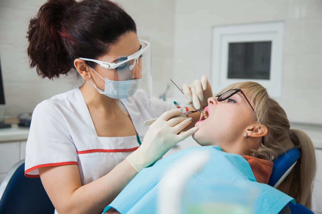
Tooth decay or cavities, also known as caries, are an everyday problem in dentistry. Cavities are the result of mineral loss caused by bacteria, and affect different parts of the tooth. One critical problem with cavities is the lack of standardisation in the way they are diagnosed. Until recently, we have lacked the necessary tools to accurately assess cavities and other common dental conditions. With caries that can be seen at the surface, such as occlusal (biting surface) caries and root caries, a diagnosis is made by simply looking into the month and by touching the tooth. Unfortunately, this directly observational mode of diagnosis is highly subjective and lacking in precision.
X-rays are used inside the oral cavity for diagnostic purposes. They represent the traditional method for imaging oral anatomy but are limited in many cases. For example, the use of bitewing radiography (X-ray) cannot be relied upon for the assessment of lesion depth. In this instance, dental professionals are left with no option but to guess how large a cavity is, based on observational evidence and their general experience. This can lead to dental professionals opting to use the drill unnecessarily.
Over the last 20 years, researchers have been investigating new tools for use in dentistry. Mr Yihua Zhu and a group of researchers at the Department of Preventive and Restorative Dental Sciences, University of California, are dedicated to advancing this cause. Mr Zhu has built upon existing research in the field of dental imaging by introducing important new methods. In particular, the group has built upon a method called short wavelength infrared (SWIR) reflectance and introduced dual techniques through the addition of transillumination imaging employing a newly invented probe.
These two methods are usually utilised independently, but Mr Zhu and his PhD supervisor, Professor Daniel Fried, have invented a way of combining the two modalities. The group has recently further refined this new tool by investigating SWIR using different wavelengths of light and comparing the results with those of standard methods in dentistry. In addition, Mr Zhu and his fellow researchers have researched the method of cross-polarisation optical coherence tomography (CP–OCT) for its significance in diagnosing dental caries.
New Tools and Techniques in Dental Diagnostics
The combination of X-rays and visual assessment in dental diagnoses has been the traditional method of choice. They provide a lot of valuable information and are routinely used in practice today. Even so, radiographs can be of limited value for the clinical assessment of certain conditions, such as root and occlusal caries. In addition, radiographs can only detect caries with a mineral loss between 30–60%, making them incapable of detecting early caries. Visual assessment is also limited in the face of common dental problems like staining. It is nearly impossible to tell non-carious staining apart from carious lesions with visual assessment, so overtreatment is also a concern.
According to the data, around one-third of patients who routinely visit the dentist at any one time have a questionable lesion (perhaps a crack or a cavity) on a tooth situated toward the back of the mouth. Dental practitioners desperately need new ways of measuring the depth of cavities and checking whether tooth decay has reached down to the dentin of the tooth. Dentin or dentine comprises the majority of the tooth and is the material lying beneath the enamel that protects the nerves. It is typically a yellowish colour and is softer than enamel (though harder than bone). The accurate assessment of conditions that have reached this part of the tooth is essential, particularly as problems in this region can lead to the need for surgery.
Two decades ago, the novel imaging methods SWIR and near-infrared (NIR) imaging became available. The coating of the tooth, otherwise known as the enamel, is highly transparent when longer wavelengths of light (short-wave infrared) are used. The tooth can be imaged from the biting surface (or the occlusal surface) when a SWIR light is shone at or below the gum line. This method is known as occlusal transillumination.
Researchers quickly saw that this optical imaging technique offered new possibilities for the early diagnosis of dental caries and for measuring the severity of cavities. Single wavelength SWIR imaging is an efficient and non-invasive technique that does not utilise ionising radiation. SWIR light is useful for detecting areas of early demineralisation (where plaque has worn away the enamel) on the tooth, so it can detect caries before they become too severe to be treated conservatively with options including fluoride varnish to promote remineralisation.
There are pros and cons to different methods and so, the prospect of combining modalities was an obvious and important part of the research agenda. Due to SWIR imaging’s high sensitivity, the increase in sensitivity of single wavelength SWIR imaging creates the problem of false positives such as glares and hypomineralisation that can be mistaken as dental decays.
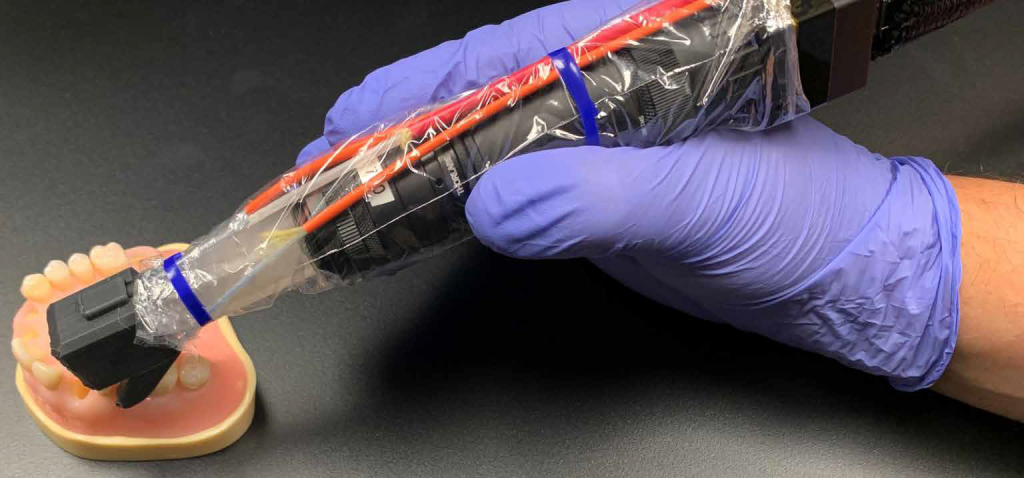
3-D printed SWIR Probe. Credit: Yihua Zhu
A Dual Approach
Mr Zhu and colleagues began looking at SWIR transillumination in combination with reflectance imaging in 2021. Transillumination uses transmitted light at a wavelength in which enamel is transparent. In reflective imaging, a wavelength with high water absorption was chosen as it generates the highest contrast of demineralisation, and light is directed at the tooth and the reflected light rays are then collected by a camera. The California group developed a new probe that would enable both methods to be used almost simultaneously. The group has tested the probe’s efficacy with extracted teeth, and they are currently testing the probe on live human subjects.
The use of dual methods offered the group the prospect of differentiating between shallow and deep cavities or lesions. When Mr Zhu and Professor Fried directed SWIR light below the crown (the part that protrudes out of the gums and bone) of the tooth while taking pictures of the biting surface, they were able to produce images with much greater levels of contrast when compared to traditional X-ray pictures. This enabled the detection of early cavities between the teeth.
This early study demonstrated that it was indeed possible to combine both the reflective and transillumination methods within a single device (or probe). That meant practitioners would no longer need to switch between devices thus saving time and improving efficiency.
More recently, Mr Zhu and the group have investigated more potential of the dual imaging probe. Altering the wavelength is important to resolving problems like staining, and stains do not absorb light at longer wavelengths. So, the application of the SWIR technique is critical to avoiding interference from staining. This would enable dental practitioners to distinguish between this problem and that of demineralisation.
Mr Zhu and his colleagues discovered that the depth of dental cavities could be correctly measured between adjacent teeth with SWIR imaging, allowing accurate assessment of lesion severity. Once again, the researchers worked with extracted teeth. The samples were collected from San Francisco Bay (where the probe was originally invented) and from Geneva in Switzerland. There were 36 teeth altogether that on further investigation revealed a total of 67 cavities.
The researchers found that their new SWIR technique using multispectral imaging was also optimal for detecting cavities on occlusal and interproximal (between adjacent teeth) surfaces. Here, it was found to be much better than traditional methods. These results were not explained by the thickness of the enamel but rather, by the probe’s placement. The method also enhanced the prospect of accurately measuring dental cavities between adjacent teeth when the probe is placed directly above the tooth biting surface. Meanwhile, the reflectance component of the dual probing method was not found to be useful for visualising cavities between adjacent teeth on tooth surfaces when the probe was directly placed facing either the cheek or the tongue side. Here only 7 out of the total 67 cavities were visible – a surprising result for Mr Zhu and the group.
In addition to enhancing diagnostic prospects, another advantage of the new tool is that it negates the need to use ionising radiation. Beyond the immediate benefit, this is also advantageous in enabling researchers to research the progression of problems like caries over time (something that cannot be justified when X-radiation is involved).
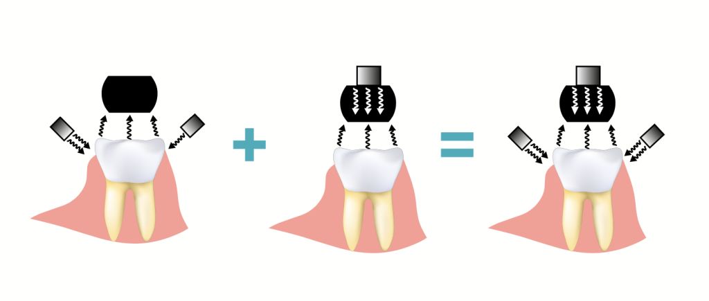
Illustration of the SWIR probe concept. Credit: Yihua Zhu
Cross-Polarisation Optical Coherence Tomography
Mr Zhu and the group have also recently investigated another tool known as CP-OCT, a non-invasive method that uses polarised light to take 3D images of the tooth. The method enhances contrast and enables imaging of the cross-sectional anatomy of the tooth. CP-OCT has proven useful in the detection of root and coronal (cracks that reach the inner quarter of the dentine) caries, loss of cementum (the calcified layer covering the root) and cases of severe demineralisation at the root surface, among other normally hidden problems.
Importantly, the high resolution brought by CP-OCT allows the detection of the transparent remineralised surface zone on top of dental caries. Remineralisation of caries indicates the start of the natural healing process of the tooth (much like scabs on top of a skin wound). Detection of a remineralised surface zone indicates that the lesion has begun this process and treatment is no longer necessary. However, the remineralised surface zone is transparent under visible light, and it is also too thin to be detected by radiographs. The only way to identify its presence is by looking at the extracted tooth and examining its histology.
But with the help of CP-OCT, Mr Zhu and the group were able to identify the presence of remineralised surface zone in live human subjects and track its growth over time. This further opens the possibility of conservative treatment in dentistry by detecting ‘arrested’ caries and overcoming the need to mechanically intervene. As we can see, the advances by Mr Zhu and his research group represent a significant contribution towards advancing modern dentistry and in particular, dental diagnostics.
SHARE
DOWNLOAD E-BOOK
CHINESE E-BOOK
REFERENCE
https://doi.org/10.33548/SCIENTIA908
MEET THE RESEARCHER

Mr Yihua Zhu
University of California
San Francisco, CA
USA
Mr Yihua Zhu is currently studying for a PhD in Oral and Craniofacial Sciences at the University of California, where he is a member of Professor Daniel Fried’s research group. Mr Zhu completed a Bachelor of Arts in Physics at the University of Southern California in 2016, followed by a Master’s degree in Medical Physics at the University of Pennsylvania in 2018, accumulating valuable research experience in various research groups along the way. He is a member of the American Association of Physicists in Medicine and also the International Society for Optics and Photonics. Mr Zhu has published in several international, peer-reviewed journals and presented his work at large conferences dedicated to advances in dentistry.
CONTACT
E: Yihua.Zhu@ucsf.edu
W: https://dentistry.ucsf.edu/about/students/yihua-zhu
T: @YihuaZ_
KEY COLLABORATORS
Professor Daniel Fried (PhD supervisor, PhD thesis mentor)
Professor Cynthia Darling (PhD thesis mentor)
Professor Donald Curtis (Clinical Collaborator)
FUNDING
National Institute of Dental and Craniofacial Research/National Institutes of Health
FURTHER READING
Y Zhu, M Kim, D Curtis, et al., Active Surveillance of Root Caries in Vivo with CP-OCT, Diagnostics, 2023, 13(3), 465. DOI: https://doi.org/10.3390/diagnostics13030465
Y Zhu, D Fried, Measurement of the Depth of Lesions on Proximal Surfaces with SWIR Multispectral Transillumination and Reflectance Imaging, Diagnostics, 2022, 12(3), 597. DOI: https://doi.org/10.3390/diagnostics12030597
Y Zhu, M Abdelaziz, J Simon, et al., Dual short wavelength infrared transillumination/reflectance mode imaging for caries detection, Journal of Biomedical Optics, 2021, 26(4), 043004. DOI: https://doi.org/10.1117/1.JBO.26.4.043004
REPUBLISH OUR ARTICLES
We encourage all formats of sharing and republishing of our articles. Whether you want to host on your website, publication or blog, we welcome this. Find out more
Creative Commons Licence (CC BY 4.0)
This work is licensed under a Creative Commons Attribution 4.0 International License. 
What does this mean?
Share: You can copy and redistribute the material in any medium or format
Adapt: You can change, and build upon the material for any purpose, even commercially.
Credit: You must give appropriate credit, provide a link to the license, and indicate if changes were made.
SUBSCRIBE NOW
Follow Us
MORE ARTICLES YOU MAY LIKE
Dr Norio Mitsuhashi | Measuring Respiratory Motion to Improve Precision in Lung Radiation Therapy
Dr Norio Mitsuhashi, former Professor of the Department of Radiation Oncology at Tokyo Women’s Medical University, leads revolutionary clinical research into optimising stereotactic body radiation therapy for lung cancer. Dr Mitsuhashi and his colleagues examine whether routinely available patient and tumour characteristics can predict respiratory tumour motion, a critical source of uncertainty in high precision radiotherapy. Their findings suggest that respiratory motion cannot be reliably inferred, and must instead be measured directly in every patient.

Professor Terry C. Hrubec | Clean is good – but is too clean better?
Quaternary ammonium compounds are a large class of compounds used as disinfectants in hospitals, restaurants, healthcare and animal care facilities, and are popular as household cleaners. With disease outbreaks increasing our fears about infections, the use of disinfectants has skyrocketed in recent years. Understandably, we all want to feel safe. However, as Professor Terry Hrubec from the Department of Biomedical Sciences of E. Via College of Osteopathic Medicine discovered, such products may be causing more harm than good.
Professor Abraham P. Lee | Delivering Cancer Immunotherapy with Acoustic-Electric Precision, AESOP’s Fact not Fable
Chimeric Antigen Receptor (CAR) T-cell therapy offers life-saving potential, particularly against blood cancers, but severe side effects such as cytokine release syndrome (CRS) limit its safety. These toxicities are linked to uncontrolled CAR expression levels on the T-cell surface. Led by Professor Abraham P. Lee, researchers at the University of California, Irvine, have developed an advanced microfluidic system, called the Acoustic-Electric Shear Orbiting Poration (AESOP) platform, to precisely control the dose of genetic material delivered into primary T cells. This innovation promises safer, more homogeneous, and highly effective cellular immunotherapies.
Dr Ray Stewart | Barriers to Dental Care for People with Special Needs: A Crisis of Neglect and Inaction
For people with special healthcare needs, something as basic as visiting a dentist can be nearly impossible. A ground-breaking paper by researchers at the University of California, San Francisco (UCSF) exposes the scale of this crisis. By outlining potential paths forward, Dr Ray Stewart and Dr Ben Meisel offer hope for significant improvements in access to essential dental care.