
The brain’s ability to manage stress and guide behaviour, including making decisions or interacting with others, relies in part on an area called the medial prefrontal cortex. But exactly how this region controls the body’s internal responses has remained unclear. New research on mice led by Prof Hong-Wei Dong and his team at the University of California Los Angeles (UCLA) sheds light on a little-studied part of the brain that may play a key role.
The work reveals a complex network, unconvering a previously undefined “primary visceromotor cortex” in the mouse brain, that helps link thoughts, feelings, and physical state. This discovery could reshape our understanding of how the brain controls stress, emotion, and internal bodily functions, and offer new insights into human mental health disorders.
Understanding the Brain–Body Connection
Most of us are familiar with the idea that the brain controls movement—reaching for a glass, walking across a room, speaking to a friend. But equally vital, and much less understood, is how the brain controls the invisible actions of the body: regulating heart rate, hormone levels, digestion, and the fight-or-flight response to stress. This internal regulation, known as visceromotor control, ensures our body remains in balance—a state scientists call homeostasis.
The parts of the brain responsible for this kind of control, particularly in the prefrontal cortex, have long been a subject of speculation. While we’ve known that some brain regions influence things like hormone release or autonomic function, the idea of a dedicated cortical region acting as a command centre for internal bodily control has remained elusive.
Now, in a landmark study, Dr Dong and his team have provided the first comprehensive map of such a system in the mouse brain—identifying what they call the “primary visceromotor cortex.” This discovery offers not only a fresh framework for understanding how the brain integrates bodily and emotional states, but also promises new avenues for studying disorders like depression, post-traumetic stress disorder (PTSD), and anxiety.
Mapping the mouse brain
The work was led by a multidisciplinary neuroscience team aiming to untangle the complex circuitry of the medial prefrontal cortex (MPF, which is also known as mPFC)—a brain region involved in emotional regulation, decision-making, and responses to stress. Their goal was to define the architecture and function of the MPF more precisely and understand how it might govern internal bodily processes.
The researchers suspected that hidden within the MPF lay a structured network responsible for controlling neuroendocrine (hormonal) and autonomic (involuntary nervous system) functions. But without a detailed wiring diagram of the region, this was hard to prove. So, using a combination of whole-brain connectivity mapping, functional experiments, and molecular profiling, the team set out to chart this previously unexplored territory.

Discovering the Primary Visceromotor Cortex
The team focused on two subregions of the MPF: i) the dorsal peduncular cortex (DP), which is further subdivided into the superficial layer (DPs) and deep layer (DPd), and ii) the ILA (infralimbic area). These areas had previously been loosely associated with stress and autonomic control, but the team’s high-resolution mapping revealed something more striking: these three areas form a functional core that connects directly to brain regions responsible for controlling hormone release, heart rate, digestion, and respiration.
They named this trio the “primary visceromotor cortex”—in analogy to the primary motor cortex, which commands voluntary movement. In a similar way, this newly identified visceromotor system sends direct projections to areas like the hypothalamus and brainstem that manage internal body states.
How Each Subregion Plays a Unique Role
Prof Dong and colleagues discovered that different parts of the visceromotor cortex each have a specialised function:
- The DPd is closely connected to a brain region called the paraventricular nucleus of the hypothalamus (PVH), which helps trigger the release of stress hormones. This suggests that the DPd plays a central role in how the body responds to stress at the hormonal level.
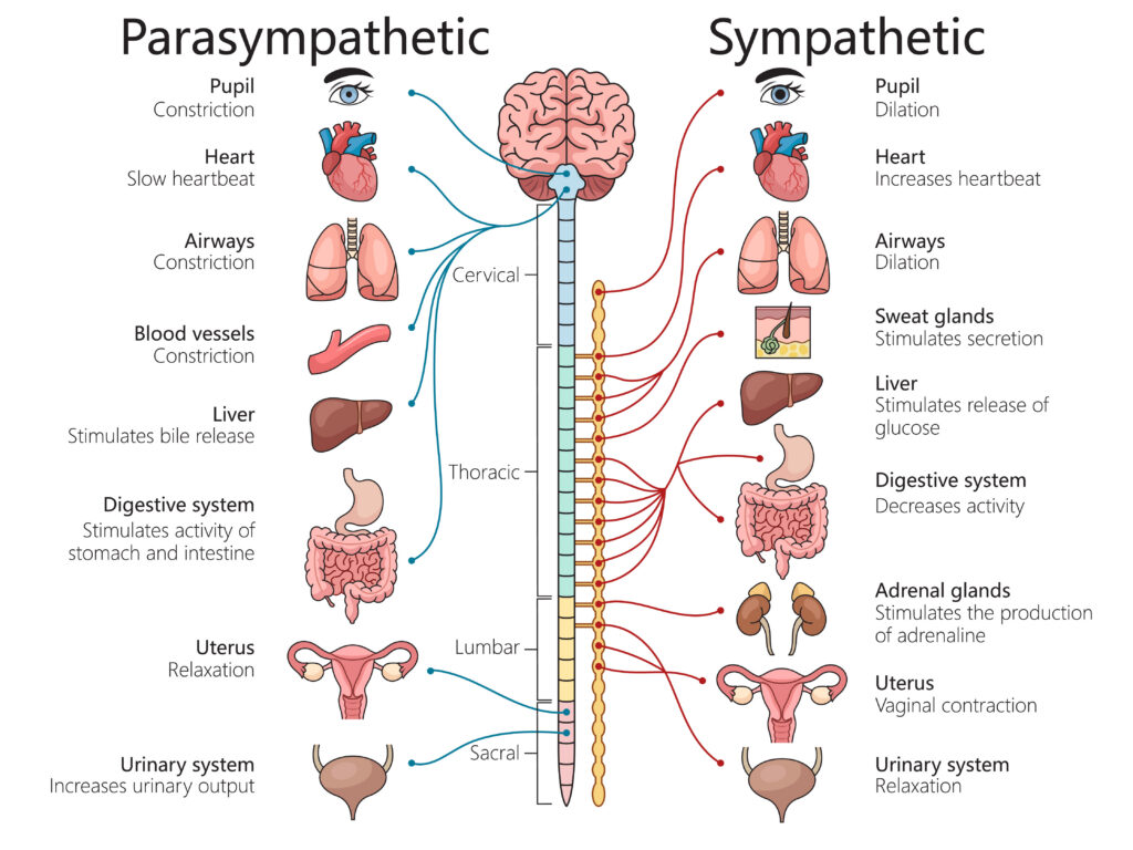
- The DPs, on the other hand, sends signals to areas that activate the body’s sympathetic nervous system—the system that prepares us for action in stressful situations, often called the “fight-or-flight” response. It also dampens activity in the parasympathetic nervous system, which normally helps the body relax and recover.
- In contrast, the infralimbic area (ILA) supports calming, parasympathetic functions. It also helps regulate emotional responses and supports social behaviour by influencing other brain regions involved in mood and interaction.
These findings show that different areas within the MPF have specific, top-down control over how the body reacts—whether that’s ramping up for action or slowing down to rest and recover.

Building the Bigger Picture: A Unified Network Model
Looking beyond individual areas, the research team also mapped out the wider network of brain regions connected to the MPF. At the centre of this system is the DP region, which acts as a hub, gathering and processing information from across the brain. These inputs include:
- smell and pheromone signals,
- emotion and memory centres (like the amygdala and hippocampus),
- sound and sensory processing areas, and
- the insular cortex, which helps track internal body signals like heartbeat and gut feelings.
Once this information is processed, the DP region passes it along to the visceromotor cortex. This helps the body prepare for whatever is happening—whether it’s facing a threat, interacting socially, or dealing with emotional stress.
The MPF also connects to parts of the brain that control movement. This means it can link how we feel to how we act—for example, by triggering actions like fleeing, freezing, or approaching others. In this way, the MPF helps the brain turn what we sense and evaluate into appropriate behavioural and bodily responses in real time.
What This Means for Mental Health
One of the most compelling aspects of the study is its relevance to human health. While the research was conducted in mice, the MPF regions identified—particularly DPd, DPs, and ILA—are thought to correspond to parts of the ventromedial prefrontal cortex (vmPFC) in humans. This brain area has long been implicated in mood disorders, but results have been inconsistent.
The researchers suggest that the functional differences between neighbouring MPF regions may explain these inconsistencies. For example, while DPd appears to promote stress hormone release, ILA may suppress it. Treating them as a single region may have obscured these distinct roles in past studies.
This new mapping offers a clearer framework for investigating depression, anxiety, PTSD, and even the biological basis of social behaviour and emotional regulation.

Looking Ahead: From Brain Maps to Therapies
Prof Dong and team now hope to expand their research to better understand how this visceromotor system functions in real-world contexts—such as during stress, social interaction, or illness. They are also exploring how sex differences and early life experiences may shape these circuits and contribute to individual differences in stress reactivity.
Ultimately, this foundational brain map could inform the development of more targeted treatments for psychiatric and stress-related disorders. By understanding which parts of the brain control internal body states, scientists may one day be able to fine-tune brain circuits to support emotional resilience, social functioning, and physical health.
SHARE
DOWNLOAD E-BOOK

REFERENCE
https://doi.org/10.33548/SCIENTIA1321
MEET THE RESEARCHER

Professor Hong-Wei Dong
University of California, Los Angeles (UCLA), Department of Neurobiology, 36-120 CHS, Los Angeles, CA 90095-1763
Professor Hong-Wei Dong is a renowned neuroanatomist and Professor of Neurobiology at University of California, Los Angeles (UCLA). He obtained his MD and PhD in Neuroscience in Xi’an, China, before completing doctoral and postdoctoral training in neuroanatomy at the University of Southern California (USC). In 2004, he joined the Allen Institute for Brain Science in Seattle, where he was one of the first to work on the Allen Brain Atlas (ABA) and he went on to create the original Allen Reference Atlas (Dong, 2007, Wiley)—now a foundational resource for large-scale brain mapping projects worldwide.
In 2006, Prof Dong launched the Mouse Connectome Project at UCLA, pioneering mesoscale connectomics. He later served as Associate Professor at USC, securing over $40 million in NIH BRAIN Initiative funding, before returning to UCLA in 2020 as Professor of Neurobiology and founding Director of the UCLA Brain Research and Artificial Intelligence Nexus (B.R.A.I.N.). A global leader in neuroanatomy, he co-leads the NIH BRAIN INITIATIVE Cell Census Network (BICCN) Anatomy and Morphology Working Group and has published extensively in high-impact journals. Prof Dong is also a committed educator, teaching brain architecture across all academic levels at UCLA.
CONTACT
W: https://neurobio.ucla.edu/people/hong-wei-dong-md-phd
KEY COLLABORATORS
Weizhe Hong, Department of Biological Chemistry & Department of Neurobiology, David Geffen School of Medicine at UCLA, University of California Los Angeles, Los Angeles, CA, USA
Peyman Golshani, Department of Neurology, David Geffen School of Medicine at UCLA, University of California Los Angeles, Los Angeles, CA, USA
Carlos Cepeda, IDDRC, Jane and Terry Semel Institute for Neuroscience and Human Behavior, Department of Psychiatry and Biobehavioral Sciences, David Geffen School of Medicine at UCLA, University of California Los Angeles, Los Angeles, CA, USA
Paul Micevych, Department of Neurobiology, David Geffen School of Medicine at UCLA, University of California Los Angeles, Los Angeles, CA, USA
FUNDING
NIH grants U01MH114829 (H.-W.D.), R01 MH094360-06 (H.-W.D.), 1R01NS133744-01 (H.-W.D.)
FURTHER READING
H Hintiryan, M Zhu, P Zhao, et al., Neural networks of the mouse visceromotor cortex, Nature, 2025. DOI: https://doi.org/10.1038/s41586-025-09360-w
![]()

REPUBLISH OUR ARTICLES
We encourage all formats of sharing and republishing of our articles. Whether you want to host on your website, publication or blog, we welcome this. Find out more
Creative Commons Licence (CC BY 4.0)
This work is licensed under a Creative Commons Attribution 4.0 International License. 
What does this mean?
Share: You can copy and redistribute the material in any medium or format
Adapt: You can change, and build upon the material for any purpose, even commercially.
Credit: You must give appropriate credit, provide a link to the license, and indicate if changes were made.
SUBSCRIBE NOW
Follow Us
MORE ARTICLES YOU MAY LIKE

Dr. Colleen Fisher | Singing to the Lions: Tackling Childhood Trauma in Zimbabwe Through Creative Psychosocial Support
Mental health challenges are one of the biggest issues facing children and adolescents across the world, particularly in low-resource settings. In Zimbabwe, a 3-day intervention called Singing to the Lions is helping young people build resilience through creative, traumainformed workshops. Dr Colleen Fisher and colleagues in Zimbabwe and the U.S. led the first formal evaluation of the intervention, offering new insight into how locally grounded intervention approaches can support youth mental health at scale.
Professor Neil Coffee – Professor Vincent Versace | Mapping Health Access: Using Address-Level Intelligence for Smarter Services
Accessing healthcare is a serious challenge for people living in rural and remote Australia. Large distances, sparse populations, and limited services can prevent residents from receiving care when they need it. Professors Neil Coffee and Vincent Versace at Deakin University’s Centre for Australian Research into Access (CARA) are leading research to model healthcare service access across the country, to provide new insights that can guide health planning and policy, as well as other services such as education. This work combines the curation of detailed address level residential dwellings and road network data to calculate access to service metrics (time and distance). These metrics are applied to the simulated residential dwelling population, to quantify the population with poor access to health services.

Professor Tony Gerard Butler | Mental Health Treatment: A Critical Pathway in the Criminal Justice System
Professor Tony Butler of the University of New South Wales explores the dynamic relationship between mental health and justice, unravelling the transformative potential of mental health treatment in the criminal justice system. By navigating the complexities of mental health interventions and their implications for reoffending rates, Professor Butler’s research provides valuable insights into the inherent challenges – and opportunities – found at the intersection of mental health and justice.

Dr David Hansen – Abhishek Juneja | Coming of Age: Active Balancing in Adolescence
Adolescence, the transitional stage between childhood and adulthood, comes with challenges as well opportunities for growth and learning.