
Dr Björn Schönnagel – Foetal Magnetic Resonance Imaging and the Diagnosis of Congenital Heart Defects: A New Approach
Foetal magnetic resonance imaging (MRI) is increasingly used for the imaging of many types of medical conditions and research continues into its use. However, MRI of the foetal cardiovascular system has encountered significant difficulties due to the constant movement caused by the beating of the heart which results in blurring of images. Dr Björn Schönnagel and his team of the University Medical Centre Hamburg-Eppendorf, Germany, have recently introduced a newly developed Doppler ultrasound (DUS) device that is compatible with MRI and allows registration of the foetal heartbeat in utero. This information is mandatory to synchronise the cardiac movement with MR image acquisition, and therefore allows successful high-quality imaging of the human foetal heart.
Congenital heart disease (CHD) represents one of the most common birth defects in foetuses with no chromosomal abnormalities and is a leading cause of infant mortality and sickness. Currently, the incidence of CHD is 6–9 per 1,000 live births. Monumental breakthroughs in surgery and diagnostics have been achieved in the past century, leading to an increased survival rate for newborns with severe CHD. As such, over 85% of babies born with a CHD now live into adulthood. However, the accurate diagnosis of CHD in the prenatal stage is often crucial for successful short- as well as long-term treatment – and ultimately survival. Critically, the development of diagnostic imaging modalities is likely to be invaluable for medical teams in supporting parents and informing the planning of postnatal care.
Existing Approaches in Foetal Heart Imaging
Currently, echocardiography (that is, ultrasound of the heart) is used to create images of the foetal heart during pregnancy. During this procedure, a small probe is placed on the mother’s abdomen, which sends out ultrasonic sound waves at a high frequency. When the probe is placed in certain locations and at certain angles, the ultrasonic sound waves are reflected by the mother’s and baby’s tissues (also the baby’s heart), depending on their reflection properties. The reflected waves are recorded and displayed as an image, thus providing images of the foetal heart which are used for diagnostic purposes.
However, not all cases of CHD can easily be detected by echocardiography. Especially in the later stages of pregnancy echocardiography can be limited as a technique due to the position of the foetus, calcification or hardening of the foetal bones, maternal obesity, and the reduction of the volume of amniotic fluid. This means that new techniques could be helpful in improving the accurate detection and classification of heart defects during this later phase of pregnancy.
E-book

Reference
https://doi.org/10.33548/SCIENTIA348
Share
‘We believe that this novel technique of direct foetal cardiac gating is a promising approach in the prenatal evaluation of the foetal cardiovascular system, especially in cases where echocardiography is limited or inconsistent.’
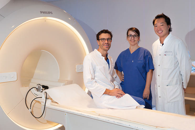
The foetal MR imaging team: Björn Schönnagel (left), Dr Manuela Tavares de Sousa (middle), Professor Jin Yamamura (right). Credit Björn Schönnagel.
Magnetic resonance imaging (MRI) is a medical imaging technique that uses a strong magnetic field and radio waves to generate ultrafast images of the organs of the body. To date, MRI has been used for the imaging of many different foetal organs, especially the central nervous system. In many ways it can be superior to ultrasound, e.g., imaging planes can be easily acquired in any anatomical orientation, not being dependent on the acoustic window like ultrasound, and image quality is not affected by issues such as foetal positioning, interference of bone and only in a lesser extent to maternal obesity. The performance, accuracy, and quality of MRI improve with gestational age, in contrast to foetal ultrasound, which becomes progressively more difficult. In addition, MRI can also provide accurate functional information, such as blood flow characteristics.
However, MRI of the foetal cardiovascular system has been hampered due to a number of technical difficulties. The greatest concern is that due to the constant movement caused by the beating of the foetal heart, blurring of images occurs if the image acquisition and the cardiac cycle are not precisely synchronised. In the adult patient, synchronisation of the heartbeat and magnetic resonance image acquisition is achieved by triggering the heartbeat (known as cardiac gating) using conventional electrocardiogram (ECG; with surface electrodes on the patients’ chest wall) or pulse oximetry. However, due to the inaccessible position of the foetus in utero, assessment of ECG or pulse oximetry information to directly trigger the foetal heartbeat is impossible.
With the help of physical engineers Dr Fabian Kording, Dr Christian Ruprecht, and Kai Fehrs, the foetal imaging team of Dr Björn Schönnagel, Professor Jin Yamamura and Dr Manuela Tavares de Sousa of the University Medical Centre Hamburg-Eppendorf, Germany, have recently developed an MR-compatible Doppler ultrasound (DUS) device that allows successful foetal cardiac gating for high-quality MRI of the human foetal heart. The developed DUS device is based on the principles of a cardiotocogram, which is used in pregnancy to monitor foetal heart rate and uterine contractions. The probe of a cardiotocogram is placed on the abdomen of the pregnant woman and uses Doppler ultrasonic sound waves for detecting both the foetal heartbeat and uterine contractions. The scientists modified this conventional probe by replacing several parts of the probe to make it compatible for use in high magnetic field environments such as MRI. In a series of experiments, they have been able to confirm the effectiveness of this technique for foetal cardiac gating.
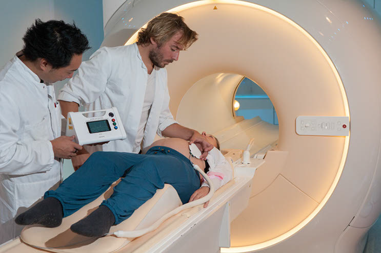
Positioning of the MR-compatible Doppler ultrasound device before foetal MRI examination. Credit Björn Schönnagel.
Early Developmental Work
A foetal sheep animal model was the first important step in the development of the MR-compatible DUS device. Together with the MR-compatible DUS probe dedicated software components were developed for optimisation of foetal heartbeat recognition. To demonstrate the effectiveness of the MR-compatible DUS device, the researcher team performed foetal heart triggering in five foetal sheep as part of their early developmental work. The scientists placed the DUS probe on the abdomen of the pregnant sheep. When a constant DUS signal (that is, a foetal heartbeat) was received, the probe was fixed in place by a belt. In this way, DUS signals were translated into trigger signals by the dedicated software and trigger pulses were conducted to the MR system to allow synchronisation of MR image acquisition with the foetal heartbeat.
 Efficacy in Human Foetal Assessment
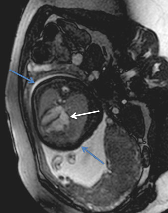
In an initial study, fifteen human foetuses at late gestational age were examined using the new MR-compatible DUS device for foetal cardiac gating. The DUS probe was fixed on the maternal abdomen to generate trigger signals from foetal cardiac motion during the MR examination. The triggering signals and image quality were analysed, demonstrating reliable trigger signals and high image quality of the dynamic images of the foetal hearts.
Dr Schönnagel and colleagues also looked at the feasibility of foetal phase-contrast-MR angiography, which is a functional MRI method for characterisation of blood flow. The study investigated blood flow haemodynamics of the descending aorta, the largest artery in the human body, in third-trimester human foetuses. Using their DUS device for foetal cardiac gating, they compared foetal blood flow rate assessed by MRI with the reference standard of Doppler ultrasound measurements of blood flow. They found that their MR-compatible DUS device for foetal cardiac gating allows for foetal MR angiography in the foetal descending aorta revealing high correlation with Doppler ultrasound measurements.
The group also used their DUS device for foetal cardiac triggering with MRI to see if they could successfully differentiate normal foetal hearts from foetuses with CHD in comparison to foetal echocardiography as the reference standard. The researchers looked at eight foetuses with a normal heart and four with a CHD. All foetuses were in the third trimester of pregnancy. A variety of anatomical landmarks and quantitative measurements of the foetal heart were assessed for comparison. Direct cardiac gating using the DUS device allowed continuous triggering of the foetal heart. Foetal cardiac MRI and echocardiography revealed an overall consistency of both quantitative and qualitative measures for differentiation of foetuses with normal hearts and foetuses with CHD.
For the first time, Dr Schönnagel and his group were able to show that dynamic foetal cardiac MRI using external cardiac triggering allows diagnosis of CHD and that furthermore, this was in similar in effectiveness to the reference standard of foetal echocardiography. The researchers note the importance of this finding, commenting ‘We believe that this novel technique of direct foetal cardiac gating is a promising approach in the prenatal evaluation of the foetal cardiovascular system, especially in cases where echocardiography is limited or inconsistent.’
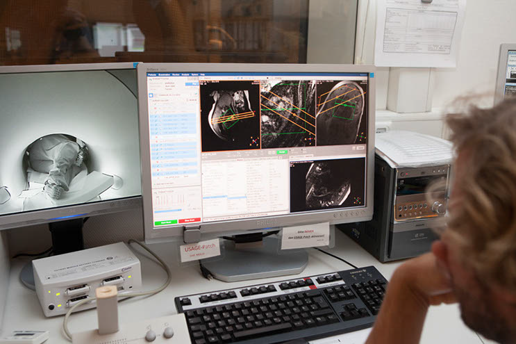
Planning an acquisition of MR images using the MR-compatible Doppler ultrasound device. Credit Björn Schönnagel.
The Future of Direct Foetal Cardiac Gating
Drawbacks still remain in the use of MRI for foetal cardiovascular imaging. More specifically, the imaging plane needs to be defined before imaging begins and as such, is vulnerable to the effects of foetal movement. Additionally, the foetal cardiac trigger signal may be lost due to foetal movement, necessitating replacement of the DUS probe on the maternal abdomen. The start-up company ‘northh medical’ is currently on the way to advance and manufacture the DUS device for widespread usage.
However, despite these difficulties, MRI has the exciting potential to aid in the evaluation of foetal CHDs, especially in later-stage pregnancies where foetuses may not be adequately evaluated by Doppler ultrasound alone. The ability to trigger the foetal heart could potentially be a significant technological advance in foetal cardiovascular MRI, aiding in the diagnosis of abnormal foetal heart structure and blood flow in pregnancies where ultrasound is not possible or where the findings of which are inconclusive. Using MRI paired with the novel device, Dr Schönnagel and his team may provide a viable alternative to foetal ultrasound in the future, offering better imaging of the foetal cardiovascular system and consequently, diagnosis of CHD worldwide.
Meet the researcher

Dr Björn Schönnagel
University Medical Center Hamburg-Eppendorf
Hamburg
Germany
Dr Björn Schönnagel completed his medical degree in 2009 at Georg August University Göttingen and is now a senior physician in the Department of Diagnostic and Interventional Radiology and Nuclear Medicine at the University Medical Centre in Hamburg, Germany, where he has worked since 2009. Dr Schönnagel is an active researcher as well as practitioner, and a co-founder of ‘northh medical GmbH,’ the company responsible for the development of the MR-compatible Doppler ultrasound device.
CONTACT
W: https://www.uke.de/english/physicians-and-scientists/arztprofilseite_bj%C3%B6rn_sch%C3%B6nnagel.html and http://northh-medical.com/
KEY COLLABORATORS
Manuela Tavares de Sousa, MD, Department of Obstetrics and Fetal Medicine
Jin Yamamura, MD, Department of Radiology
Jochen Herrmann, MD, Department of Pediatric Radiology
Carsten Rickers, MD, Department of Pediatric Cardiology
Fabian Kording, PhD, Kai Fehrs, PhD, and Christian Ruprecht PhD, northh medical GmbH
FUNDING
German Research Foundation
FURTHER READING
F Kording, et al, Doppler ultrasound triggering for cardiovascular MRI at 3T in a healthy volunteer study, Magnetic Resonance in Medical Sciences, 2017, 16, 98–108.
M Tavares de Sousa, et al, Dynamic fetal cardiac magnetic resonance four chamber view imaging using Doppler ultrasound gating in the normal fetal heart and in congenital heart disease: comparison to fetal echocardiography, 2018, Ultrasound in Obstetrics and Gynecology, doi: 10.1002/uog.20167.
F Kording, et al, Dynamic fetal cardiovascular magnetic resonance imaging using Doppler ultrasound gating, 2018, Journal of Cardiovascular Magnetic Resonance, doi: 10.1186/s12968-018-0440-4.
BP Schoennagel, et al, Fetal blood flow velocimetry by phase-contrast MRI using a new triggering method and comparison with Doppler ultrasound in a sheep model: a pilot study, Magnetic Resonance Materials in Physics, Biology and Medicine, 2014, 27, 237–244.
BP Schoennagel, et al, Fetal dynamic phase-contrast MR angiography using ultrasound gating and comparison with Doppler ultrasound measurements, 2019, European Radiology, doi: 10.1007/s00330-018-5940-y.