Dr Kazuhiro Yasufuku – Getting Really, Really Small to Treat Lung Cancer

Feb 11, 2017Medical & Health Sciences

Thoracic surgeon and research scientist Dr Kazuhiro Yasufuku and his colleagues at the University of Toronto in Canada aim to use the latest in nanoparticle and high technology to treat lung cancer without major surgery.

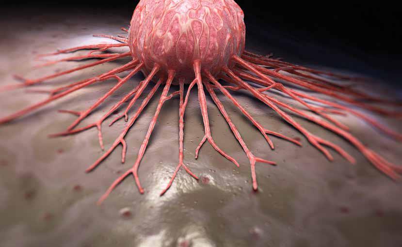
Lung Cancer is not Just Any Cancer

Cancer is scary enough, but lung cancer – cancer arising in the lungs and airways – is one of the most distressing diseases imaginable, and with good reason. While lung cancer is not always the most common malignancy, depending on where you live, it is certainly one of the deadliest. In Great Britain, for example, lung cancer ranks below breast cancer and prostate cancer in incidence, but according to Cancer Research UK, it is the most common cause of cancer death. More than 35,000 people died from lung cancer in the UK in 2012. Cancer Australia reports that lung cancer is the fifth most common cancer in that country, yet it is the leading cause of cancer death, with almost 9,000 deaths due to lung cancer yearly. Things are dire in Canada, where Lung Cancer Canada claims that lung cancer is both the most common cancer and more people die of lung cancer than breast cancer, colorectal cancer and prostate cancer combined – with almost 21,000 people dead in 2015. Similarly, the United States Centres for Disease Control documents that more Americans die from lung cancer than any other type of cancer – 157,423 in 2012. No matter where you live, lung cancer is deadly, so it makes sense that medical researchers are keenly interested in new and better ways to treat it.
As a surgeon trained in operations on the chest, Dr Kazuhiro Yasufuku knows how difficult traditional lung cancer treatments can be. For one thing, the chest is well protected by a bony cage of bone and cartilage, making traditional chest surgery challenging for the patient. Incisions through the chest muscles, spreading or even breaking ribs and cutting or stretching cartilage – all this is physically demanding and can lead to long convalescences and pain for the patient. It is also technically challenging to remove tumours that may be close to vital structures, like the heart and large arteries and veins. Of course, you may also be taking out pieces of the lung, or even an entire lung. And non-surgical treatments for lung cancer are not all peaches and cream. Radiation beams aimed at a tumour in the lung can also do significant collateral damage to adjacent normal tissue. Chemotherapy can affect the whole patient. After all, chemotherapy is simply using toxic substances to try to kill the tumour before poisoning the patient. Dr Yasufuku knew there must be a better way.

How About Making Tiny Holes?

In earlier days of surgery, many surgeons found ways to avoid large incisions, turning to various lighted scopes to perform ‘minimally invasive surgery’. Perhaps first to jump on this band wagon were the gynaecologists, who eschewed a long, midline incision (or even the ‘bikini cut’ transverse incision) in favour of small puncture wounds through which they could insert laparoscopy equipment. Several incisions no larger than a centimetre could be placed to allow lighted scopes and manipulators to be used to perform significant surgical operations on women. This surgery was often termed ‘band aid surgery’, because all you had covering your small incisions when you went home were band aids. General surgeons soon got on board, avoiding a long incision under the right ribs to take out a gall bladder, for example, and replacing it with a few small poke holes through which a laparoscopic cholecystectomy could be accomplished. Now, hernia repair, colon resection, appendectomy and other major surgical procedures can be accomplished through the laparoscope. Similarly, minimally invasive surgery for lung cancer became available via Video Assisted Thoracoscopic Surgery (VATS) in the 1990s. However the adoption by thoracic surgeons has been slow due to counter-intuitive hand movements to manipulate the instruments and 2-dimensional images with limited magnification. In order to solve these problems, Dr Yasufuku along with colleagues at Toronto General Hospital made history in Canada in 2012.
In October of 2012, Dr Yasufuku used a da Vinci robotic surgical apparatus – multiple remotely controlled arms manipulating instruments placed through small incisions in the patient’s body – to remove part of the lung of a 78-year old man with lung cancer. Instead of suffering through a 20- to 30-centimetre thoracotomy incision, along with spreading apart of ribs and the attendant post-operative pain, the patient had four small 1-centimetre incisions in his chest that left almost no scarring. In fact, the one ‘big’ incision that was necessary was a 3-centimetre incision only needed so as to extract the excised portion of the lung. The patient was up and eating breakfast the next day and discharged from hospital soon after in virtually no pain or discomfort. At follow-up visit he was cancer free. Since that first surgery, Dr Yasufuku has performed many other lung cancer surgeries using this minimally invasive technique. Now, not only patients with, say, gynaecologic problems can benefit from band aid surgery. But this minimally invasive lung cancer procedure solves only one problem, that of a large and painful incision. There is still the question of how to recognise and localise tumours before you cut into – or at least poke holes in – the patient’s chest. To be able to place the multiple small incisions for minimally invasive surgery, the surgeon needs a precise location of the tumour. Besides that, what if the tumour is too close to vital structures to excise, or the patient is too fragile for even minimally invasive surgery? Is there a way to get to the tumour and localise it better with our current diagnostic methods? Better than that, can you get to the tumour and destroy it without classic surgery at all? Dr Yasufuku had ideas about this.

Photothermal ablation

Endoscopic excitation of dissociated porphysomes using navigation bronchoscopy
Bronchoscopic fluorescence visualization by porphysome of the target tumor
Bronchoscopic laser photothermal ablation

First, Getting into the Chest Without a Scalpel

One way to get into a patient’s chest without going between or through the ribs is to use the doorway Mother Nature gave us. Certainly pulmonologists use bronchoscopes all the time – flexible tubes that allow the physician to visualise the inside of the trachea and bronchi, almost all the way down into the alveoli themselves. Dr Yasufuku and fellow researchers considered this a great avenue down which to pursue lung tumours. The only problem is how do you actually see the tumour when your view is inside the airway tubes? In other words, if your lighted tube and camera is inside a bronchus, how do you visualise a tumour outside the bronchus, out in the lung tissue itself?
Because of modern medical imaging techniques and more astute medical strategies, doctors are now seeing increasing number of patients with small, non-palpable lung tumours that are extremely difficult to biopsy or find during surgery. Surgical resection is the standard of care and provides the best survival benefit for patients with early stage lung cancer, but if you cannot find the tumour, you cannot remove it. As well, some patients are not physically fit enough for surgery. That is where Dr Yasufuku and his group are getting really creative and jumping into the world of nanoparticles and looking at the manipulation of matter on a molecular scale. Dr Yasufuku and his group in Toronto have been developing innovative methods to solve these problems by application of new nanotechnology, the porphysome and the ICG lyposome, to their minimally invasive surgery techniques. Both the porphysome and the ICG lyposome were developed at the University of Toronto.

Porphysome? What’s a Porphysome?

The porphysome is a first-of-its-kind, multifunctional, all-organic lipid nanoparticle discovered by Dr Yasufuku’s colleague Dr Gang Zheng. The traditional role of lipids in this arena has been limited mainly to the use of liposomes – spherical nanoparticles consisting of a lipid bilayer – to deliver chemotherapy drugs. The clinical success of these liposomes has been based on their high biocompatibility, biodegradation and clearance properties, as well as their high therapeutic efficacy or contrast enhancement capabilities. The organic characteristics of lipids allow them to avoid eliciting an undesired immune response and help guarantee safe excretion from the body. A number of drug-loaded liposomes have been approved or are in clinical trial for various cancers. Unfortunately, the role of these lipid preparations is simply that of a passive carrier used to increase the circulation time of chemotherapy drugs and prevent interaction of the drugs with blood cells.

But now, with the synthesis of porphyrinlipid – a porphyrin structure conjugated to a phospholipid – and the discovery of porphysome nanovesicles by Dr Zheng, the role of phospholipids in supramolecular structures has shifted from a simple support to an intrinsic imaging and therapeutic agent. Porphyrins are a group of aromatic organic compounds that are often pigmented – the word porphyrin comes from the Greek word for purple. One well-known porphyrin is heme, the pigmented portion of red blood cells and component of haemoglobin. And

Intraoperative localization

Thoracoscopic excitation of dissolved porphysomes in the target tumor
Thoracoscopic fluorescence visualization of the target tumor
Minimally Invasive limited resection under fluorescence guidance

The use of nanoparticles, particularly porphysomes and ICG lyposome, is a quantum leap in our ability to fight lung cancer.

because of the complex molecular structure of porphyrins, they can be used for a variety of purposes, including even carrying a metal ion attached to the porphyrin molecule by chelation. So a combination of porphyrin molecules of varying design with phospholipids – essentially tucking a specially designed porphyrin into a liposome – gives cancer researchers the ability to do wonderful things on a molecular scale. Porphysomes can be designed to accumulate into tumours and to emit bright fluorescence and near-infrared (laser) light to preferentially heat the tumour tissue. Porphysomes can be built containing metal molecules to allow for intraoperative localisation of early-stage lung cancer during minimally invasive surgery. Properly designed porphysomes could allow ultra-minimally invasive photothermal therapy of lung cancer in patients that are not surgical candidates. Porphysomes light up the tumour so the surgeon can snake a laser down a bronchoscope and zap it without making an incision at all. The possibilities are endless for Dr Yasufuku and his team.

Add ICG Lyposome and Start Working

Beyond the fascinating porphysome, the Toronto researchers also designed the ICG lyposome. This is a novel lipid-based liposomal nanoparticle encapsulating the commercially available fluorescent dye indocyanine green plus an X-ray contrast agent. This combination would allow precise imaging using both CT and near-infrared techniques to help guide the surgeon during minimally invasive thoracic surgery. The ability to ‘see’ even very small tumours, either during surgery or during bronchoscopy, would be a great advantage for surgeons like Dr Yasufuku. And with these two exciting nanoparticle additions to their armamentarium, the group at Toronto is planning a couple of studies to use both porphysomes and ICG lyposome to fight lung cancer.First, Dr Yasufuku has a grant to construct an image-guided localisation platform for minimally invasive lung surgery. He plans to develop a new system in which the surgeon would get intraoperative real-time information on blood vessel anatomy as well as tumour location in the lung by lighting up the tumour with ICG lyposome. That way the tumour will show up on CT and near-infrared imaging so the surgeon can do his minimally invasive magic with precision. This project includes putting the ICG lyposome together with the minimally invasive techniques and various scanning devices and perfecting it in preclinical animal models for ultimate use on human cancer patients. In other words, use animals first to make sure the nanoparticles, the X-ray machines and the surgical system all coordinate and everything runs without a hitch.
Perhaps even more exciting, Dr Yasufuku wants to skip the surgery altogether and work on an ‘ultra-minimally invasive’ treatment for lung cancer, even less invasive than his band aid surgery. He wants to use infrared light delivered to the target tumour though a bronchoscope to destroy the malignant tissue by local heating, i.e. photothermal therapy. Specially designed porphysomes will be used to localise the target tumour by fluorescence imaging and to enhance the local absorption of the laser light, something porphyrins are good at. The project also includes perfecting the technique in preclinical animal models and then moving into human clinical trials, since it is something that has never been done in humans. The first-in-human trials will be performed in the Guided Therapeutic Operating Room (GTx OR) built in the Toronto General Hospital. The project team in Toronto already has all the expertise: Dr Yasufuku knows how to wield a bronchoscope, his colleague Dr Brian Wilson is an expert in fluorescence endoscopy, Dr Robert Weersink can work the X-ray machines, Dr Zheng wrote the book on the nanoparticles and Drs Wilson and Weersink are all about photothermal laser ablation. This team has been working together for years now and nobody else is more prepared to take this giant leap into the world of nanoparticles and minimalist surgery to defeat a major killer, lung cancer.

Meet the researcher

Dr Kazuhiro Yasufuku, MD, PhD
Associate Professor,
Department of Surgery,
University of Toronto, Canada

Dr Kazuhiro Yasufuku is a thoracic surgeon and research scientist. He received his MD in 1992 from the School of Medicine of Chiba University, Japan. In 2002 he also received a PhD from the Graduate School of Medicine there. After a residency in Thoracic Surgery in Japan and fellowships in Thoracic Surgery and Transplantation in both Canada and Japan, Dr Yasufuku moved to Toronto, where he is currently the Director of Endoscopy at the University Health Network (UHN) as well as the Director of the Interventional Thoracic Surgery Program at the Division of Thoracic Surgery, University of Toronto. He is also the clinical lead of Thoracic Surgery within the Guided Therapeutics program at UHN and leads the Thoracic Robotic Surgery Program at UHN. He is a Scientist at the Latner Thoracic Surgery Research Laboratory, Toronto General Research Institute, UHN. He has authored or co-authored over 150 peer-reviewed journal articles, as well as book chapters and other publications.

CONTACT

KEY COLLABORATORS

Dr Gang Zheng: Professor of Medical Biophysics, University of Toronto Senior Scientist, Ontario Cancer Institute
Dr Brian Wilson: Professor and Head of Medical Biophysics, University of Toronto Senior Scientist, Ontario Cancer Institute
Dr Robert Weersink: Assistant Professor, Radiation Oncology, University of Toronto Affiliated Faculty, Techna Institute for the Advancement of Technology for Health (Techna)
Dr David Jaffray: Senior Scientist, Princess Margaret Cancer Centre Executive VP Technology and Innovation, University Health Network Director, Techna Institute, Techna Institute for the Advancement of Technology for Health (Techna)
Dr Jinzi Zheng: Assistant Professor, Institute of Biomaterials and Biomedical Engineering, University of Toronto Scientist, Techna Institute for the Advancement of Technology for Health (Techna)
Dr Shaf Keshavjee: Professor, Division of Thoracic Surgery & Institute of Biomaterials and Biomedical Engineering Vice Chair, Innovation, Department of Surgery, University of Toronto
Dr Ming Tsao: Professor, Department of Laboratory Medicine and Pathobiology, University of Toronto Senior Scientist, Ontario Cancer Institute, Division of Applied Molecular Oncology

FUNDING

Canadian Cancer Society
Canadian Institutes of Health Research
Natural Sciences and Engineering Research Council of Canada
Ontario Institute of Cancer
The Physicians’ Services Incorporated Foundation
Olympus Medical Systems Inc.
Intuitive Surgical Inc.
Novadaq Corp.
Veran Medical

REFERENCES

Yasufuku K, Pierre A, Darling G, de Perrot M, Waddell T, Johnston M, da Cunha Santos G, Geddie W, Boerner S, Le LW, Keshavjee S, J Thorac Cardiovasc Surg., 2011, 142, 1393–1400.

Nakajima T, Zamel R, Anayama T, Kimura H, Yoshino I, Keshavjee S, Yasufuku K, Ann Thorac Surg., 2012, 94, 2097–101.

Anayama T, Nakajima T, Dunne M, Zheng J, Allen C, Driscoll B, Vines D, Keshavjee S, Jaffray D, Yasufuku K, PLoS One, 2013, 8, e67355.

Wada H, Hirohashi K, Anayama T, Nakajima T, Kato T, Chan HH, Qiu J, Daly M, Weersink R, Jaffray DA, Irish JC, Waddell TK, Keshavjee S, Yoshino I, Yasufuku K, PLoS One, 2015, 10, e0126945.

Nakajima T, Geddie W, Anayama T, Ko HM, da Cunha Santos G, Boerner S, Wang T, Wang YH, Li M, Pham NA, Tsao MS, Yasufuku K, Lung Cancer, 2015, 89, 110–4.