
Dr. Sharon Ruthstein – The Importance Of Copper In The Human Cell
Dr. Sharon Ruthstein researches the biological pathways involving metal ions, especially copper, by using Electron Paramagnetic Resonance (EPR) Spectroscopy. Here, she discusses the importance of copper in the human body and the implications for future diagnostic and therapeutic care.
What is your educational and research background and what triggered your interest in research of the biological pathways involving metal ions?
When I did my PhD at the Weizmann Institute in Israel, under the supervision of Prof. Daniella Goldfarb, I acquired my expertise in EPR Spectroscopy. After graduation, I started my postdoctoral work at the University of Pittsburgh (PA) under the supervision of Prof. Sunil Saxena who is interested in the coordination of copper ions in various proteins. Here, I noticed that many proteins contain metal ions. I thought it would be interesting to use these paramagnetic metal ions in EPR spectroscopy instead of the commonly used site-directed spin labelling method, to resolve biological questions.
What is the importance of metals, such as copper, in living organisms?
Metal ions are involved in a variety of important biological and chemical processes at in the cell, including oxygen transport, biosynthesis, electron transfer and drug metabolism. Copper, similar to iron, is involved in electron transfer reactions and oxygen metabolism. Copper is also crucial for the development of the central nervous system. Metal ions are a double-edged sword. Metal ions act as cofactors for catalysis in various enzymes but are also highly toxic. So, precise monitoring of the concentration of each metal is critical to the cell.
 If we look at copper within living organisms, what do you think is the most important step in the copper cycle that we need to understand?
To date, it is known that there are three different pathways involved in the copper cycle in human cells: to cytochrome C, to SOD1, and to the Golgi apparatus. I believe that the most important pathway is the transfer mechanism of copper to the copper transporter “Atp7b” of the Golgi apparatus via the copper transporter “Ctr1” and through the copper chaperone “Atox1”. This cycle controls the copper concentration within the cell and is therefore of tremendous importance for the copper regulation. Mutations in the copper transporter Atp7b were found to be responsible for Menkes and Wilson disease.
Why does the localisation of copper within the cell need to be controlled so carefully?
High concentrations of copper can be deleterious, leading to oxidative damage of proteins, lipids and nucleic acids. The coordination of copper has been linked to promoting peptide aggregation which results in forming amyloid plaques characteristic for Alzheimer’s, Parkinson’s and prion disease. On the other hand, copper deficiency is just as dangerous. This results in metabolic abnormalities due to a decreased function of copper dependent proteins.
Disruption of the copper homeostasis can eventually lead to neurological disorders. So, it is of vital importance to understand every step of the human copper cycle. With this knowledge, we can build a fundamental understanding of possible disruptive elements to the copper homeostasis.
At the moment, you are working on different research projects in your lab such as developing highly sensitive detection tools. When we look at clinical practice, what do you hope to contribute?
We are developing copper biomarkers for hypoxic tissues, tissues that lack oxygen supply. For this, we use detailed knowledge of the pathway and reaction mechanism or a compound. We hope that our compound will either replace the currently used biologically active tracer molecule in PET/SPECT imaging (FDG), or complement it so our knowledge of tumour size and type will improve leading to superior diagnostic examination in cancer patients.
I have also read a research proposal in which you hope to introduce a new treatment for ALS. What do you hope to achieve with this research to alleviate the burden of ALS?
I strongly believe that our lack of knowledge of the origin and causes of amyloidgenetic diseases like Alzheimer’s disease, Parkinson’s disease and ALS is the reason for lacking good therapeutic solutions. In my opinion, because these diseases are all related to high copper concentrations, shedding light on the copper cycle will result in more effective therapeutic approaches. Possible new therapeutic options could be the use of competitive metal ions as well as gene therapy, a growing field nowadays.
EXPLORING THE BIOLOGICAL PATHWAYS OF COPPER IN THE HUMAN CELL
Metal ions are involved in many important processes in the human cell. However, little is known about the regulation of these metal ions at cellular level. Dr. Sharon Ruthstein of the University of Bar-Ilan researches the biological pathways of metal ions, especially copper, by using Electron Paramagnetic Resonance (EPR) Spectroscopy.
Metal ions
Metals are cofactors in many biological and chemical reactions in human cells, including oxygen transport and drug metabolism. More than 30% of all proteins in the cell use one or more metals to perform their specific functions and over 40% of all enzymes contain metals. On the other hand, metal ions can be highly toxic when free in biological fluids. So, a regulatory system is in place in the human body to manage the concentrations and types of metal ions in and outside the cell. Despite these regulatory systems, diseases such as Menkes, Wilson’s, Alzheimer’s, Parkinson’s and Prion which originate from metal binding to proteins, still emerge.
The importance of copper
Copper is an important metal ion in the human body. It is involved in electron transfer reactions and oxygen metabolism. Copper is also crucial for the development of the central nervous system. High concentrations of copper can be dangerous, because it leads to oxidative damage of proteins, lipids and nucleic acids. Copper deficiency is just as dangerous resulting in abnormalities in the metabolism.
Copper accumulates in the human body through diet. After a change in its atomic structure, copper is delivered to the cell by the copper transporter called “Ctr1”. After arrival in the cell, specific copper chaperones are responsible for delivering the copper to specific cellular pathways. Dr. Sharon Ruthstein’s lab focusses on the main copper transporter “Ctr1”, on the “Atox1” copper chaperone and the “Atp7A and Atp7B” copper transporters. Mutations in “Atp7A” and “Atp7B” are found to be the leading cause for Menkes and Wilson’s disease. The “Atox1” copper chaperone and the “Ctr1, Atp7A and B” copper transporters are proteins that directly interact in a copper stimulated-manner.
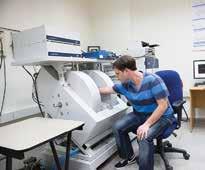
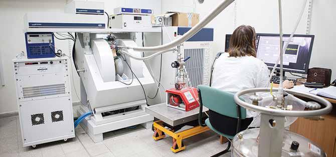
EPR Spectroscopy in copper research
The main tool in the research of metal ions used in Dr. Ruthstein’s lab is continuous wave (CW) and pulsed electron paramagnetic resonance (EPR) spectroscopy. Over the last decade, EPR spectroscopy has emerged as an important method for resolving structure-function relationships in proteins. The power of this tool lies in the sensitivity to both atomic level changes and nanoscale fluctuations. EPR can characterize certain properties of the protein’s functional state as well as information of its dynamics. EPR can also probe small fluctuations in the protein’s structure. And, more importantly, measurements can be conducted in conditions that closely mimic the natural environment of the protein. EPR does not require crystallization and it is not limited to the protein’s size. EPR is the perfect tool to target complex biological systems.
Dr. Ruthstein uses EPR spectroscopy together with site-directed spin labelling techniques to explore structural changes that occur in the methionine motif when it binds with copper. Methionine motifs are methioninerich metal binding segments present in many human proteins. These proteins are involved in the transportation of copper to other cellular pathways and in the protection of copper from oxidation. These methionine motifs bind copper as well as silver, another metal ion. Proteins that can bind different metal ions in similar way should be able to identify between them so they can shuttle them to their own pathway in the cell. Dr. Ruthstein showed that the methionine segments undergo different structural changes while binding copper instead of silver.
She also looked at the copper transporter “Ctr1” as mentioned earlier. With this study she found out that the methionine motif, the methionine-rich metal binding segments in proteins, changed it’s conformation when approaching the Ctr1 transporter. She discovered that these changes where dependent of which metal ion it was bind to, copper or silver.
 The future in EPR spectroscopy
Studying the interactions between proteins involved in the metal cycle is often difficult. For instance, a protein undergoes only minor changes in its conformation when binding to metal ions. These minor changes can only be studied with biophysical tools that have a very high resolution. High resolution tools are an important step forward in the research of the functions of proteins at molecular level. It remains however difficult to study these proteins in a living environment, called in-vivo. In living cells, the proteins interact with other proteins, nucleic acids and co-factors. It is very difficult to replicate the cellular environment outside the living cell, called in-vitro.
So, there is a tremendous need to develop a highly sensitive, high resolution biophysical tool that will be able to provide structural and molecular information on the interactions between proteins which are involved metal ion transfer in and outside the living cell.
There are two major challenges in resolving the biological pathways that involve metal transfer in the cell. Firstly, it is necessary to follow the biological pathway in its natural environment. Secondly, the biophysical tool need to be highly sensitive to even minor changes in the conformation that the proteins undergo as a consequence of metal binding or protein-protein interaction during the biological pathway. In recent years, the biophysics community has been struggling with the development of these high-sensitive tools for in-cell detection. Dr. Ruthstein is working on a new EPR and labelling methodology. This tool is based on time-resolved EPR measurements of photo-induced radical pairs (RP) and will be able to resolve the biological pathways with higher sensitivity and resolution, both in-vitro as in-cell.
Implications for clinical practice
CT and MRI imaging have long been the standard diagnostic tools in cancer research. However, CT and MRI are only limited effective in determining the metabolic and functional information of certain tissue. For this purpose, PET and SPECT are much more powerful techniques. There techniques are able to image an increasing variety of physiological phenomena because of the possibility to select a radio-tracer that specifically targets a particular mechanism. Because the incidence of cancer grows, there is continuous demand for the development of new radio-tracers for early cancer detection and chemotherapy targets.
A feature of all solid tumours is hypoxia which is the deprivation of adequate oxygen supply. The association between a tumour and low oxygen levels makes it a high priority target for cancer therapy. Dr. Ruthstein is developing a highly sensitive copper-tracer for PET measurements using the gained knowledge on the copper transfer mechanisms which can be used in diagnostic procedures in cancer. She consults with renowned oncologists in Israel and the USA to further improve its diagnostic and therapeutic options in cancer.
Meet the researcher
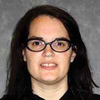
Dr. Sharon Ruthstein
Department of Chemistry
Faculty of Exact Science
University of Bar-Ilan, Israel
Dr. Sharon Ruthstein received her Ph.D. in chemistry, with honors, from the Weizmann Institute of Science in Israel. She has been recognized by the Wolf foundation with the Krill prize for excellent young scientists in 2015. She received the Auto Schwartz prize from the Weizmann Institute in 2007. Currently, she is a senior lecturer and researcher at the department of chemistry of the Bar-Ilan University where she is particularly interested in the copper homeostasis and the structure and function of proteins.
CONTACT
T: +972 3 738 4329
W: http://ch.biu.ac.il/ruthstein
W: http://www.ruthstein-lab.com/
KEY COLLABORATORS
Prof. Dan Major, Bar Ilan University, Israel
Dr. Eitan Okun, Bar Ilan University, Israel
Prof. Nir Ben-Tal, Tel-Aviv University, Israel
FUNDING
ISF, Marie-Curie, Israel Chief Scientist
REFERENCES
Meir, A.; Natan, A.; Moskovitz, Y.; Ruthstein, S.; Utilizing EPR spectroscopy to identify essential key residues for the copper transfer between CusB N-terminal domain and the metallochaperone CusF in E.coli. Metallomics, 2015, 7, 1163-1172.
Shenberger, Y.; Shimshi, A.; Ruthstein, S.; EPR spectroscopy shows that the blood carrier protein, human serum albumin, closely interacts with the N-terminal domain of the copper transporter, CTR1. J. Phys. Chem. B. 2015, 119, 4824-4830.
Shenberger, Y.; Gottlieb, H.E.; Ruthstein. S.; EPR, NMR, and CD spectroscopy provide inputs on the coordination of Cu(I) and Ag(I) to a disordered methionine segment. J. Biol. Inorg. Chem. 2015, 20, 719-727.
Levy, A.; Yarmaiyev, V.; Moskovitz, Y.; Ruthstein, S. Probing the structural flexibility of the human copper metallochaperone, Atox1 dimer and its interaction with the CTR1 c-terminal domain. J.Phys. Chem. B. 2014, 118, 5832-5842.
Shenberger, Y.; Yarmiayev, V.; Ruthstein, S.; Exploring the interaction between the human copper transporter, CTR1, c-terminal domain and a methionine motif, in the presence of Cu(I) and Ag(I) ions, using EPR spectroscopy. Mol. Phys. 2013. 111, 2980-2991.

Download