
Professor Karl Forchhammer – Awakening Sleeping Bacteria
Professor Karl Forchhammer and his colleagues analyse how cyanobacteria can survive and recover from long periods of starvation. They use the model strain Synechocystis PCC 6803, a non-diazotrophic, unicellular cyanobacterium. When deprived of a nitrogen source, the cells become chlorotic and survive in a dormant state. Upon re-addition of nitrate, a genetically determined program is initiated that brings back the cells to vegetative life.
Dormant Cyanobacteria
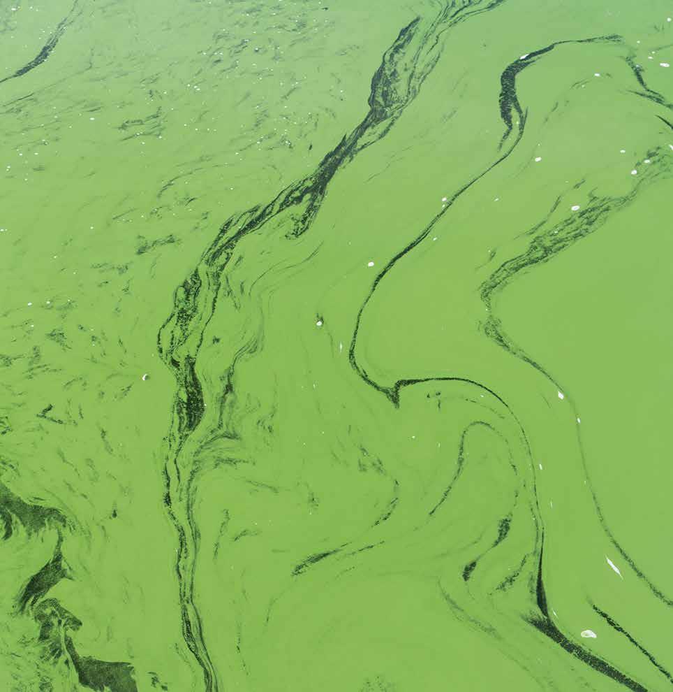
Cyanobacteria are prokaryotic cells that are also known as blue green algae. This name comes from that fact that cyanobacteria contain a blue pigment called phycocyanin, which along with green chlorophyll gives cyanobacteria a blue-green appearance. Cyanobacteria are essentially responsible for life as we know it. During the Archaean and Proterozoic Eras (2.5 billion years ago), it was cyanobacteria that were responsible for creating the Earth’s oxygen atmosphere. Prior to this, the atmosphere had a very different chemistry, one that was unsuitable to sustain life in its present form.
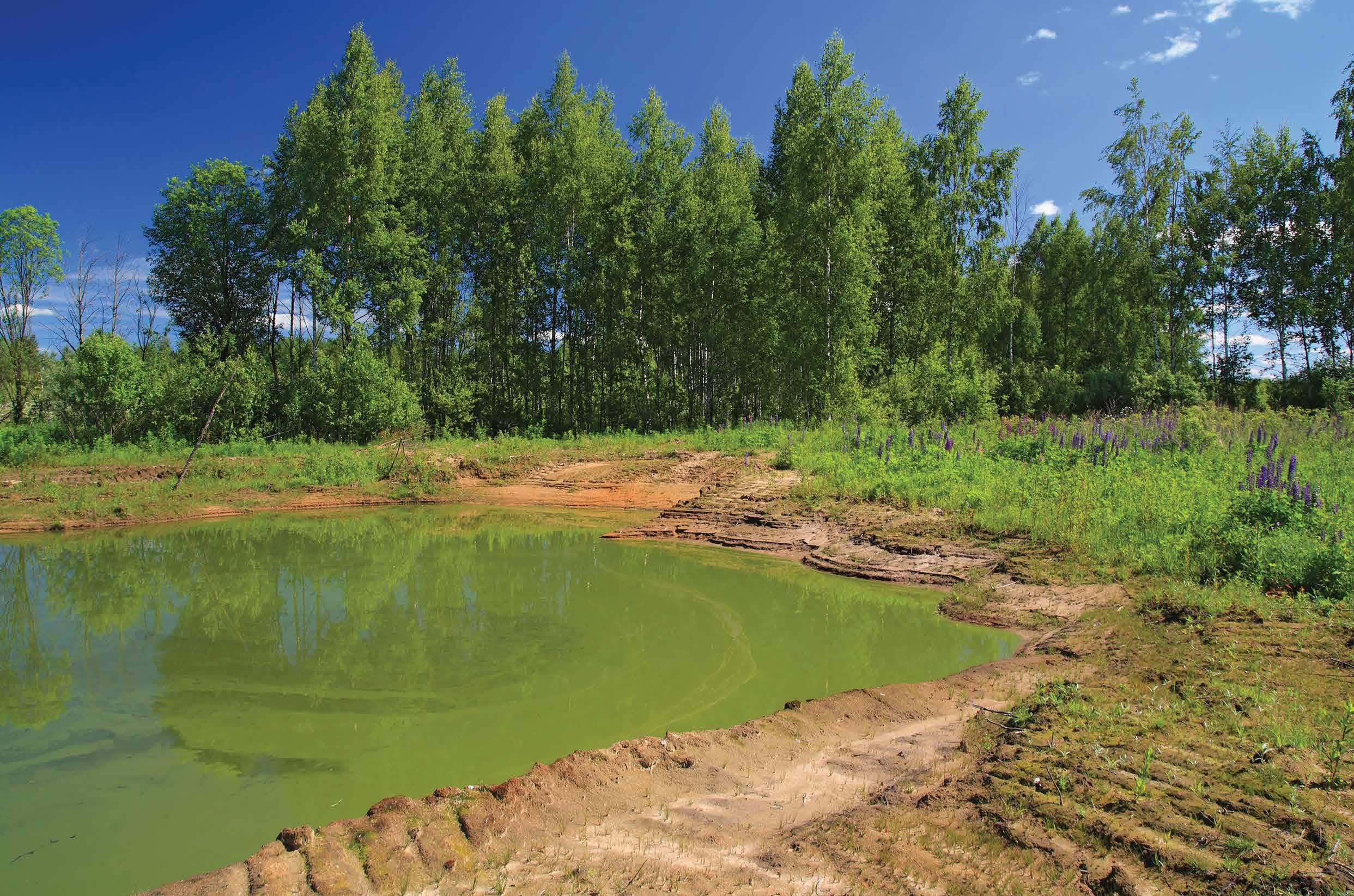
Cyanobacteria reside in a wide range of habitats that include frozen lakes, acidic bogs, deserts and volcanoes, but they are most commonly found in aquatic environments. In addition, they are found in soil, on rocks and even in the atmosphere. In the light-exposed biosphere, cyanobacteria are ubiquitous. There they function as key players in global carbon and nitrogen cycles.
Cyanobacteria are photoautotrophic organisms that absorb carbon dioxide and release oxygen. Several cyanobacteria can also absorb, or ‘fix’, elemental nitrogen present in the atmosphere, and thus, they do not have to rely on other combined nitrogen sources. In low nitrogen environments or in low carbon dioxide environments, cyanobacteria have gained to the ability to adapt and survive over the course of their evolution. These environmental challenges often limit growth in many terrestrial and aquatic ecosystems.
With respect to nitrogen use, cyanbacteria are divided into two physiological groups. The diazotrophic strains evade nitrogen starvation by expressing an enzyme called nitrogenase, which fixes all-pervading di-nitrogen (N2) gas. In contrast, the non-diazotrophic strains halt their growth when a combined nitrogen source is absent and switch their metabolism from anabolism to maintenance. To make this switch, cyanobacteria first degrade their photosynthetic pigments. This process is known as chlorosis, and during its course, the cells noticeably change in colour from bluegreen to yellow and they become dormant.
The ability of prokaryotes to become dormant is widespread, especially when nutrients in the environment are limited. There is a notion that dormant bacteria comprise a ‘seed bank’ – a reservoir of cells that can be resuscitated when favourable conditions are present again. This mechanism is largely responsible for the fitness of bacterial populations and contributes to the spreading of bacterial pathogens. In different bacterial taxa, growth limitation activates various molecular and cellular processes allowing the survival bacterial populations. The ways in which the cells enter dormancy and recover from the dormant stage are rather diverse. Some bacteria form endospores or exospores, others create encapsulated cysts, or even generate apparently non-differentiated persister cells, which is the case in Mycobacterium tuberculosis. In order to survive these extended periods of starvation, the non-differentiated resting cells have the ability to maintain a residual metabolic activity. The ability of the bacteria to rapidly resuscitate from dormancy ensures the successful spread of bacterial populations, but this mechanism remains unclear.
To study the formation of dormant cyanobacterial cells, researchers have previously utilised the freshwater strain S. elongatus subjected to nitrogen starvation as a model. Based on these studies, researchers have determined that a protein called NblA, whose expression is controlled by the Nbl regulatory system, initiates the degradation of the major light-harvesting pigments. When the cells are moved to a nitrogen depleted medium, glycogen starts to build up. This accumulation of glycogen occurs very rapidly – within 12 hours – whereas it takes more time until the pigments are fully degraded – up to 5 days. These cells will only stop growing once they have completed a final cell division – which takes about 12 hours.
If the period of nitrogen deficiency is prolonged, the cells will degrade a majority of their cellular proteins and their photosynthetic apparatus until they reach a final chlorotic stage. When the cells enter into this stage, they sustain only residual levels of photosynthesis, which are approximately 0.1 % of their initial activity. By doing this, the cells are able to preserve full viability over at least six months.
Another unicellular cyanobacterium strain, Synechocystis sp. strain PCC 6803, has a similar response to nitrogen deprivation, and this species is widely used as a model organism to study underlying aspects of photosynthesis and cyanobacterial physiology. When these cells are in nitrogen deficient conditions, this strain produces a second carbon-storage polymer, polyhydroxybutyrate (PHB), in addition to the glycogen granules, that is presumed to contribute to the ability of the cell to handle chlorosis. In addition, some preliminary results suggested that Synechocystis can achieve long-term survival under nitrogen-deficient conditions, and these chlorotic cultures are particularly efficient at recovering from chlorosis. Given that this strain has such a rapid recovery process, it is optimal for studying the resuscitation of a dormant bacterium. By doing so, researchers can gain insights into a fundamental bacterial survival strategy. Indeed, this is precisely what Professor Karl Forchhammer and his team at the University of Tübingen have done. They have used cholortic Synechocystis cells to study the organisation of resuscitation at the cellular and molecular level as an example of bacterial awakening from dormancy.
Two Phases of Resuscitation
As part of a DFG-supported research training group, RTG 1708 (Molecular principles of bacterial survival strategies), PhD student Alexander Klotz tested the viability of Synechocystis cells and the accumulation of PHB (the carbon storage polymer) and glycogen during prolonged nitrogen depletion for up to 42 days. He demonstrated that, of the two carbon reserves, glycogen accumulated almost immediately after nitrogen was depleted. In contrast, the accumulation of PHB occurred much more slowly. In addition, almost all of the cells remained viable after being deprived of nitrogen for 42 days, and they rapidly recovered following the addition of a nitrogen source.
In addition, the research team used the longterm chlorotic cells to further understand the phases of resuscitation. When the cells were resuscitated by adding nitrate to the medium, they began to turn green again within 48 hours, and were completely pigmented and growing exponentially after 72 hours. They also found signs of photosynthetic activity after 24 hours.

As we have discussed, chlorotic cells have two types of carbon reserves – PHB and glycogen – that can fuel initial respiration. Given that previous studies suggest that PHB could be important for recovery, Professor Forchhammer and his group sought to determine whether this was true, as they had detected no significant decrease in PHB content during the first 24 hours of recovery. Additionally, they had observed that PHB degradation did not start before the cells had resumed photosynthetic activity.
In their experiments, the team actually found that PHB is not required for the recovery of chlorotic Synechocystis cells. In contrast, glycogen was immediately used after starting the resuscitation program by adding nitrate to the cultures. Based on their physiological studies, Professor Forchhammer and his group discovered two phases of the resuscitation process. In the first phase, which is within the first 16 hours after the reintroduction of nitrogen, respiration is activated, which is supported by glycogen consumption. As the cells transition to the second phase, photosynthetic activity increases at the same time that the cells start to become re-pigmented. In this second phase, despite reconstituting photosynthesis, which takes over the cellular energy supply, glycogen continues to provide cells with carbon skeletons for anabolic needs.
Structural Changes During Resuscitation
The structural changes that cells undergo when they experience different cellular and molecular processes often provide clues into the mechanisms behind such processes. Therefore, Professor Forchhammer and his team were interested in visualising the morphological changes of Synechocystis cells during their recovery. In collaboration with Dr Roman Sobotka (Trebon, Czech Republic) they found that the fully developed chlorotic cells, in contrast to the growing cells, were almost completely absent of a photosynthetic thylakoid membrane and instead were completely filled with glycogen granules. The granules disappeared during resuscitation, and 24 hours after recovery the thylakoid membranes started to reappear and were fully reconstituted by 36 hours. After 48 hours of recovery, the cells had almost completely re-established thylakoid membranes and a structure resembling lipid bodies was visible. By 66 hours the cells resembled the exponentially growing cells.
In addition, the team monitored the cell size and cellular DNA content during the recovery from chlorosis. Dr Watanabe found that in the healthy exponentially growing cells there was a narrow distribution of chromosome numbers per cell, centring at about 5–6 chromosomes per cell. In long-term chlorotic cells, the distribution was broader, with many cells having up to the double number of chromosomes per cell. Only when the cells started to divide again after having completely recovered, the chromosome number dropped again to initial values, indicating that the cell cycle was arrested in a pre-divisional stage during chlorosis.
Gene Expression Changes During Resuscitation
Microarray analyses are used to gain a deeper insight into complex processes, because they provide clues as to the genes that are regulating such processes. Professor Forchhammer’s collaborator Professor Hess (University Freiburg) designed a high-density microarray to further understand the molecular mechanism behind resuscitation in recovering cells.
During the process of resuscitation, they found that the level of 1,572 transcripts, which corresponded to 17.6% of the entire transcriptome, was significantly changed during resuscitation. Among these transcripts, 781 were increased and 791 were decreased in abundance. Based on the time course of the changes, these positively and negatively responding transcripts were classified into three major groups. These groups reflected the chronological order of the transcriptome remodelling during resuscitation.
Interestingly, mRNA from the cells in chlorosis have an overall transcript abundance that is similar to that of growing cells based on the yield of the extracted mRNA. Therefore, the reduced transcriptional activity in the cells in chlorosis might be compensated by increased mRNA stability, which is a phenomenon that has been observed in various growth-arrested microorganisms. During the fully developed chlorotic state, the translational machinery is functioning at an absolute minimum level, and these transcripts make up part of the most strongly repressed of the entire transcriptome. This finding means that most of the transcripts in the chlorotic cells are translationally inactive. In addition, the microarray results revealed that there are highly abundant functionally characterised non-coding regulatory small RNAs, which are anti-correlated to their sense mRNAs. Thus, these RNAs likely play an important regulatory role in stabilising the dormant state. In contrast, the transcript levels of the most classic global transcriptional regulators were only slightly changed compared to the levels in the growing cells. If fact, this rigid transcriptional repression of the protein biosynthesis machinery is a key feature of the starvation-induced stringent response in bacteria, and reversing this repression appear to be key to the resuscitation program.
By identifying these major cellular processes and the transcriptional dynamics during resuscitation, Professor Forchhammer and his team have revealed the existence of a highly sophisticated genetic program that guides the awakening of a dormant bacterium. There studies, ultimately, provide the framework for future exploration of targeting the regulatory mechanisms that are executing this resuscitation program.
Conclusions
Professor Forchhammer and his team present the first detailed analysis and demonstration of the series of events that regulate both the entry into bacterial dormancy and the recovery to an apparently normal, photosynthetically active, vegetative cell for the cyanobacterium Synechocystis. These findings have a wide-ranges of potential implications, which will contribute to the understanding of sporulation/ germination, bacterial persistence in infection processes and may even influence our understanding of cellular aging. These studies by Professor Forchhammer shed light into the dramatic ultrastructural changes that occur in cells during chlorosis and their subsequent resuscitation. Remarkably, the recovery process is highly coordinated and does not involve the immediate regeneration of the photosynthetic apparatus, but rather it utilises a carefully orchestrated restoration. Ultimately, Professor Forchhammer and his colleagues have defined a perfect model system, which will enable researchers to dissect these processes in detail in the future.
This study was initiated and largely performed as part of the DFGsupported research training group, RTG 1708: ‘molecular principles of bacterial survival strategies’, headed by Professor Forchhammer at the University Tübingen. Under the framework of this RTG-funding, collaboration in the international team was facilitated, boosting the success of this project.
Meet the researcher

Professor Karl Forchhammer
Chair of the Department for Microbiology
University of Tübingen
Tübingen, Germany
Professor Karl Forchhammer received his Ph.D. in Microbiology at the University Munch, where he was involved in the discovery of co-translational incorporation of the 21th amino acid selenocystein. Soon after, he went on to perform postdoctoral research at the Institut Pasteur in Paris, where he discovered the serine phosphorylation of the PII signalling protein in the cyanobacterium Synechococcus PCC 7942. Between 1994 and 1999, he was assistant professor at the University of Munich, and was then appointed as Professor of Microbiology at the Justus-Liebig-University, Giessen. Since 2007 he is the chair of the department for Microbiology/Organismic interactions at the University of Tübingen.
CONTACT
KEY COLLABORATORS
Elena Ermilova, St. Petersburg State University, Russia
Wolfgang Hess, University Freiburg, Germany
Luciano Huergo, Federal State University Curitiba, Brazil
Vicente Rubio, University Valencia, Spain
Roman Sobotka, Institute of Microbiology, Trebon, Academy of Sciences, Czech Republic
FUNDING
FUNDING Deutsche Forschungsgemeinschaft (DFG)
REFERENCES
A Klotz, J Georg, L Bucinska, S Watanabe, V Reimann, W Januszewski, R Sobotka, D Jendrossek, WR Hess, K Forchhammer, Curr. Biol., 2016, 26, 1–11.
W Hauf, K Schmid, EC Gerhardt, LF Huergo, K Forchhammer, Front. Microbiol., 2016. DOI: 10.3389/fmicb.2016.01700
K Forchhammer, J Lüddecke, FEBS J., 2016. DOI: 10.1111/febs.13584
V-R Chellamuthu, E Ermilova, T Lapina, J Lüddecke, E Minaeva, C Herrmann, M Hartmann, K Forchhammer, Cell, 2014, 159, 1188–1199.
J Lehner, S Berendt, B Dörsam, R Pérez, K Forchhammer, I Maldener, 2013. FASEB J., 2013, 27, 2293–2300.

Download