
Dr Alessia Besford | Understanding the Crown of a Nanoparticle
Medical nanoparticles are an innovative method of delivering drugs to highly specific locations in the body, such as tumours or across the blood-brain barrier. Once a nanoparticle has entered the bloodstream, it forms a crown of surrounding biomolecules called a corona. The composition of this corona depends on its biological environment and the material of the nanoparticle itself and it has significant implications for how the nanoparticle interacts with the human body. Dr Alessia Besford from the Leibniz Institute of Polymer Research Dresden studies these interactions and how they can be refined to develop more effective medical nanoparticles.
Medical Nanoparticles
Nanotechnology is a fascinating and expanding field of science that can be investigated within a multitude of fields from electrical engineering to quantum physics. It is also increasingly being utilised for medical applications including innovative imaging methods, artificial implants and targeted drug delivery. The latter is particularly exciting because the ability to send drugs to treat a very specific part of the body opens up all sorts of new, more effective treatment options known as nanomedicine.
This method uses nanoparticles, which are tiny, often synthetic, vesicles usually between 1 and 100 nm across – about 1 billionth of a metre. Nanoparticles can be made from metals, ceramics or biodegradable polymers (a larger structure made up of lots of smaller units). Whilst these materials are man-made, they are designed to break down in the body so that they can release their contents. Each particle contains a drug that has been specially designed to treat a specific disease and which works especially well when delivered to its specific target.
For example, nanoparticles can be engineered to bind only to specific cancer cells. Once a nanoparticle has been internalised by the cancer cell, it is transported to the endosome, which is acidic and acts like the cell’s stomach. As a result, the nanoparticle is dissolved and the drug inside is released and the individual cell dies, fighting the cancer whilst leaving surrounding healthy cells intact. This has a number of benefits in comparison to conventional chemotherapy treatments, including a controlled release of the drug with a specific, more effective distribution. Consequently, toxic side effects and adverse reactions are minimised in the patient.
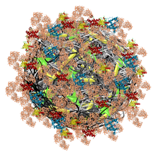
Nanoparticle with a protein corona. Credit Alessia Besford.
The Crown of Nanoparticles
Although medical nanoparticles show incredible potential, more research is needed to understand how they interact with the human body and whether these interactions could be detrimental or even toxic. This is the focus of Dr Alessia Besford’s research at the Leibniz Institute of Polymer Research Dresden in Germany, where she works together with Prof Carsten Werner at the Institute of Biofunctional Polymer Materials. She has conducted multiple studies investigating how nanoparticles interact with the human immune system. Specifically, she studies how immune cells and blood proteins interact with the nanoparticles and how the formation of the biomolecular corona, predominantly consisting of proteins, around synthetic nanoparticles affects their distribution in the body.
‘El corona’ is the Spanish word for crown and it is commonly used in science to describe crown-resembling entities. This can be a part of the body or the halo of a star. When a nanoparticle enters a biological environment (usually via the bloodstream), biomolecules like proteins and lipids adsorb onto its spherical surface, creating a corona. This crown has two layers: the ‘hard’ inner and the ‘soft’ outer biomolecular corona. The ‘soft’ layer contains more loosely bound biomolecules that can exchange with surrounding molecules, whereas the ‘hard’ layer is bound strongly and permanent.
Both the biological milieu (blood) and the surface chemistry of the nanoparticle significantly impact the biological composition of the corona. This means that how the nanoparticle is made and what it is made from are important considerations when developing a nanoparticle vehicle. Materials that reduce the ability of proteins to absorb onto the nanoparticle are called low-fouling; fouling is a term that describes the accumulation of unwanted matter on a surface. For example, poly(ethylene glycol) is often used to coat nanoparticle surfaces which results in a water layer that acts as a physical and energetic barrier against proteins.
Many studies have shown the role of the biological milieu and the surface chemistry of the nanoparticles in corona formation, although there was a lack of studies on the dynamic nature of the blood and its constant flow in the body. Therefore, Dr Besford set out to apply microfluidics to study the protein absorption of nanoparticles under flow with a number of different factors. To do this, she created an environment for her experiments that precisely mimicked the geometry of blood vessels. A variety of particles with different coatings allowed her to carry out her incubation experiments with high-, medium- and low-fouling nanoparticles. Dr Besford also tested with human blood and human plasma (blood with all cells removed) leaving a yellowy liquid instead of red).
Her results revealed that protein adsorption onto the nanoparticles is increased when they are incubated with blood compared to serum and that a dynamic flow results in a more complex corona compared to a static incubation. They also showed that as the fouling degree of the surface increases, so does the amount of different absorbed proteins. Interestingly, she found that an initial hard corona layer on the particle surface is formed 2 seconds from incubation commencement and in a second kinetic phase, a more loosely bound hard layer forms.
Dr Besford’s work here showed how design parameters and the biological environment of a nanoparticle influence the composition of its corona. Understanding corona formation can help to create more accurate drug delivery nanoparticles as predicting how they will interact in the body will affect how effective they are in vivo.

The studies of Dr Besford evolve around nanoengineered materials in human blood and how the composition of the corona affects the association of nanoparticles with human immune cells. Her aim is to predict these associations (i.e., the reactions of the human immune system) based on the composition of the corona. Credit Alessia Besford.
Investigating Nanoparticle Corona to Improve Its Medical Properties
Further work from Dr Besford identified that there are three phases of protein binding. Using an innovative technique, she mimicked the human average blood flow and utilised confocal laser scanning microscopy to observe the proteins attaching to the nanoparticles over time. In phase one, proteins irreversibly attach directly to the nanoparticle surface. In phase two, these proteins irreversibly interacted with other preabsorbed proteins. The ‘soft’ corona was formed in phase three, where proteins were bound reversibly.
This study also investigated low-fouling zwitterionic nanoparticles – particles with equal positive and negative charges to give a net zero charge. In these cases, only a soft corona formed as no proteins bound irreversibly. Through these novel approaches, Dr Besford provided a pathway for a state-of-the-art procedure for kinetics and protein fouling experiments to improve medical nanoparticle production.
Another important consideration to make when developing medical nanoparticles is how the immune system will respond to them. Lots of research has been conducted to construct low-fouling materials that also have minimal interaction with human immune cells, giving them the name stealth materials. As the nanoparticles are a foreign substance, if they are detected too readily by the immune system, they may be destroyed before they can deliver their drug load. The previous assumption was that being a low-fouling material goes hand-in-hand with being a stealth material – but Dr Besford wanted to investigate this. Her in-depth study utilising different levels of fouling materials and immune components proved that this theory is correct.
Credit Alessia Besford.
Finding the Material for Nanoparticles
Dr Besford has also dedicated research into the best materials for medical nanoparticles. One popular material is glycogen, which is a natural polysaccharide (sugar) and is therefore biodegradable and does not provoke the immune system (it has low immunogenicity). However, because it has to be synthesised to create an appropriate nanoparticle, some of these beneficial properties can be lost in the process. Therefore, along with a team led by Dr Quinn Besford (Group Leader at IPF), she developed a unique nanoparticle from oyster glycogen (OG) and polymers called PNIPAM. Excitingly, the OG-PNIPAM nanoparticles both retained the biodegradability of native glycogen and did not induce an inflammatory response from the immune system.
In one of her latest papers, Dr Besford explored spider silk-based materials due to their similarly positive elements – they are biocompatible, processable and biodegradable. The study revealed that when the silk materials are positively charged, the corona that consequently formed were made up largely of fibrinogen-base proteins. These are proteins that are necessary for blood clotting and the tests showed an increase in clotting with these positively charged particles. Comparatively, negatively-charged silk materials prevented blood clotting. The properties of the spider silk can be refined through genetic engineering, which means that a scientist could pick and choose the surface properties of the nanoparticles synthesised from it.
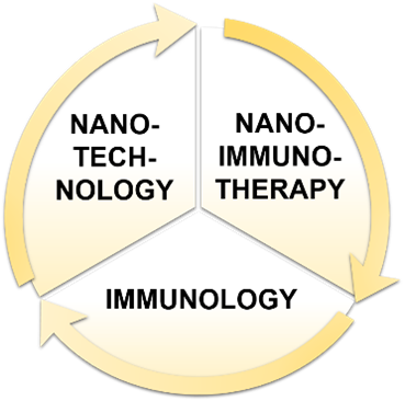
With a strong portfolio of nanoparticle research behind her, Dr Besford is continuing her work with more studies planned in the near future. In a significant stride forward from previous research, the PhD project of her student Vaidehi Londhe combines aspects of nanotechnology, nanoimmunotherapy and immunology to develop immune evasive nanoparticles.
As such, Dr Besford is continuing to build on her experience and expertise to delve deeper into the properties of nanoparticle materials, how they interact with the human body and its immune system and how collating her research can help to develop low-fouling, stealthy nanoparticles for effective medical solutions.
SHARE
DOWNLOAD E-BOOK
REFERENCE
https://doi.org/10.33548/SCIENTIA865
MEET THE RESEARCHER

Dr Alessia Besford
Leibniz Institute of Polymer Research Dresden e. V.
Germany
Dr Alessia Besford received her undergraduate degree in Polymer and Colloid Chemistry from the University of Bayreuth in Germany and stayed on to complete a Master’s degree in Polymer Science. She then left for Australia, where she achieved her PhD and completed a postdoctoral fellowship at The University of Melbourne within the ARC Centre of Excellence in Convergent Bio-Nano Science and Technology. Currently, Dr Besford is a postdoctoral research fellow at the Leibniz Institute of Polymer Research Dresden e.V. in Germany, where she works in the research group of Prof Carsten Werner at the Institute of Biofunctional Polymer Materials. She carries out research into the properties of nanoparticles and their interactions with the human immune system to develop more effective drug delivery systems.
KEY COLLABORATORS
Project group:
Dr Quinn Besford, expert in material characterisation, PhD co-supervisor
Vaidehi Londhe, PhD student
Dr Manfred Maitz, expert in hemocompatibility analyses
Dr Anna Shevchenko, mass spectrometry service leader
Recent Collaborators:
Australia
- Prof Frank Caruso, The University of Melbourne
- Prof Steven Kent, The University of Melbourne
- Prof Greg Qiao, The University of Melbourne
- Prof Edmund Crampin, The University of Melbourne
- Dr Ching-Seng Ang, The University of Melbourne
- Dr Kristian Kempe, Monash University
- Dr Francesca Cavalieri, Royal Melbourne Institute of Technology
Germany
- Prof Stefan Förster, Jülich Research Centre
- Prof Thomas Scheibel, University of Bayreuth
- Prof Andreas Fery, Leibniz Institute of Polymer Research Dresden
- Prof Carsten Werner, Leibniz Institute of Polymer Research Dresden
FUNDING
Deutsche Forschungsgemeinschaft DFG
DAAD Bayreuth-Melbourne Polymer and Colloid Network
Melbourne International Fee Remission Scholarship
Melbourne International Research Scholarship
FURTHER READING
ACG Weiss, SJ Shirbin, HG Kelly, et al., Plasma Corona Protects Human Immune Cells from Structurally Nanoengineered Antimicrobial Peptide Polymers, ACS Applied Materials & Interfaces, 2021, 13(29), 33821–33819. DOI: https://doi.org/10.1021/acsami.1c07088
QA Besford, ACG Weiss, J Schubert, et al., Protein Component of Oyster Glycogen Nanoparticles: An Anchor Point for Functionalization, ACS Applied Materials & Interfaces, 2020, 12(35), 38976–38988. DOI: https://doi.org/10.1021/acsami.0c10699
ACG Weiss, HM Herold, S Lentz, et al., Surface Modification of Spider Silk Particles to Direct Biomolecular Corona Formation, ACS Applied Materials & Interfaces, 2020, 12(22), 24635–24643. DOI: https://doi.org/10.1021/acsami.0c06344
ACG Weiss, HG Kelly, M Faria, et al., Link between Low-Fouling and Stealth: A Whole Blood Biomolecular Corona and Cellular Association Analysis on Nanoengineered Particles, ACS Nano, 2019, 13(5), 4980–4991. DOI: https://doi.org/10.1021/acsnano.9b00552
ACG Weiss, K Kempe, S Förster, F Caruso, Microfluidic Examination of the “Hard” Biomolecular Corona Formed on Engineered Particles in Different Biological Milieu, Biomacromolecules, 2018, 19(7), 2580–2594. DOI: https://doi.org/10.1021/acs.biomac.8b00196
ACG Weiss, K Krüger, QA Besford, et al., In Situ Characterization of Protein Corona Formation on Silica Microparticles Using Confocal Laser Scanning Microscopy Combined with Microfluidics, ACS Applied Materials and Interfaces, 2019, 11(2), 2459–2469. DOI: https://doi.org/10.1021/acsami.8b14307
![]()
REPUBLISH OUR ARTICLES
We encourage all formats of sharing and republishing of our articles. Whether you want to host on your website, publication or blog, we welcome this. Find out more
Creative Commons Licence (CC BY 4.0)
This work is licensed under a Creative Commons Attribution 4.0 International License. 
What does this mean?
Share: You can copy and redistribute the material in any medium or format
Adapt: You can change, and build upon the material for any purpose, even commercially.
Credit: You must give appropriate credit, provide a link to the license, and indicate if changes were made.
SUBSCRIBE NOW
Follow Us
MORE ARTICLES YOU MAY LIKE

Assoc Prof. Nicholas Brown | Rethinking Prostate Care: A New Frontier in Treating Benign Prostatic Hyperplasia
For millions of men, ageing brings with it a set of frustrating and often disruptive urinary symptoms. These symptoms, caused by benign prostatic hyperplasia, or BPH, can affect sleep, confidence, and overall quality of life. Traditionally, treatment follows a familiar path. Patients begin with medications, often for years, and may eventually progress to surgery if symptoms worsen. Yet this pathway is not without its drawbacks. Medications can cause side effects, while surgery carries risks and recovery time. In recent years, a minimally invasive interventional radiology procedure called prostate artery embolisation, or PAE, has begun to challenge this traditional model. At the forefront of this shift is a collaborative research group, led by Dr. Nicholas Brown of the University of Queensland, whose series of P-EASY studies has explored whether PAE could transform how BPH is treated, particularly at earlier stages.

Jean Lycke | Addressing Unmet Medical Needs in Mucosal Disease: A Close-to-Market Innovation Approach
Recurrent Aphthous Stomatitis (RAS) is an oral condition characterized by one or several painful mucosal ulcers. RAS affects a large proportion of the population and has a point prevalence of approximately 2–3%, daily. The etiology remains unknown, and there is currently no curative treatment. Most patients experience recurring episodes over time, with each episode typically lasting up to a week. Here, we describe the development of a mucoadhesive patch which, when applied over a RAS ulcer, provides rapid pain relief. The patch is easy for patients to apply when symptoms begin and has the potential to be used as an over-the-counter product. The development of the Mucocort mucoadhesive patch is an example of a Close-to-Market innovation strategy that embraces simplicity within a complex healthcare system. By simplifying the product concept, the team has reduced the number of regulatory steps required before market approval. This MedTech/Pharma innovation model, known as the “4R” framework – Re-purposing, Re-formulation, Re-positioning, and Re-patenting – has guided the program from concept to commercialization. In addition to the biodegradable mucoadhesive patch developed for RAS ulcers, the team is extending the innovation concept to a mucoadhesive gel formulation for the prevention and treatment of chemotherapy-induced mucositis. This gel-based program is being commercialized separately through MucoShield.
The Translational Asian Agerelated Macular Degeneration Program Phase 2 (TAAP-2): Reimagining the Future of Vision Care
Age-related macular degeneration, often abbreviated as AMD, is one of the leading causes of vision loss among older adults worldwide. In Asia, where populations are ageing rapidly, its impact is particularly profound. For many, the disease quietly erodes central vision, making everyday activities such as reading, driving, and recognising faces increasingly difficult. Against this backdrop, the Translational Asian Age-related Macular Degeneration Programme, or TAAP for short, has emerged as a bold and ambitious effort to confront the disease headon. Now in its second phase, TAAP-2 represents a significant evolution in both scientific scope and clinical ambition.
Ms. Aikaterini Dritsoula | Looking Beyond Snoring: How Hidden Airway Problems Shape Children’s Sleep
For many parents, a child’s snoring may seem harmless, even endearing. Yet in some cases, it signals something more serious. Obstructive sleep apnoea is a condition in which a child’s breathing is repeatedly disrupted during sleep. These interruptions can affect growth, behaviour, and learning. Children with this condition may toss and turn at night, struggle to concentrate during the day, or show signs of hyperactivity and fatigue. Traditionally, enlarged tonsils and adenoids have been seen as the main culprits. Surgery to remove them has long been considered the standard treatment. However, research led by Consultant ENT Surgeon Ms. Aikaterini Dritsoula of The Leeds Teaching Hospitals NHS Trust invites us to look deeper. Her work suggests that the story is often more complex, especially in very young children.