
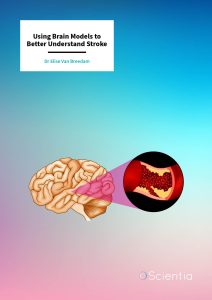
Dr Elise Van Breedam | Using Brain Models to Better Understand Stroke
Stroke is a leading cause of disability and death. Despite extensive research and the huge burden on patients and healthcare systems, treatments are sadly limited and new treatments are slow to emerge. To better understand why so many promising drugs that have worked in animal and laboratory studies failed to show the same benefits in human clinical trials, Dr Elise Van Breedam from the University of Antwerp reviewed current research methods. Her review points to the potential for human-based 3D models to bridge the gap between animal studies and human patients.
Prevalence and Consequences
Strokes are a leading cause of disability and death around the world. In Europe alone, there are over a million incidents of stroke each year, and this is likely to increase in the future as our population ages. Strokes occur when the blood supply to part of the brain is cut off, causing a wide range of symptoms depending on which part of the brain is affected.
Common symptoms following stroke include difficulty in speaking, weakness or loss of coordination, and severe headaches. People who survive are often left with long-term problems due to the damage to their brains.
The majority of strokes are ischaemic, meaning they are caused by a blood clot in a blood vessel, reducing (or even blocking) the blood flow to part of the brain. Within minutes of reduced blood flow, the brain cells in this area begin to die due to the lack of oxygen and nutrients. The initially damaged area of the brain is known as the ischaemic core. Damage to cells here is severe and likely to be irreversible. The cells in the ischaemic penumbra surrounding the core are usually less severely damaged and, with quick treatment, may recover.
Despite extensive research to date, stroke treatment is currently limited to drugs which break down blood clots or the surgical removal of the blockage. These treatments need to be administered as soon as possible after the stroke. However, due to the rapid nature of stroke damage and the limited time frame in which these therapies are actually effective, only a very small proportion of ischaemic stroke patients can be treated in this way. This leaves thousands of stroke patients without viable treatment options.
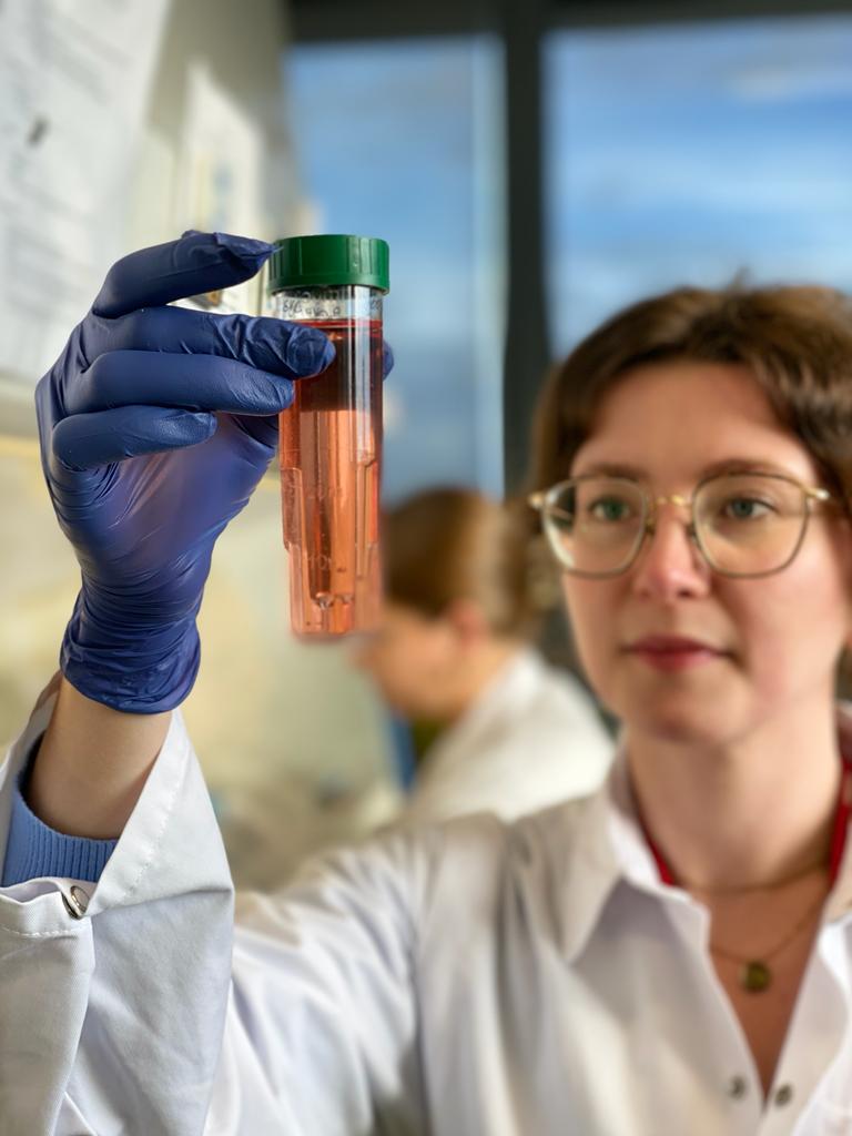
Dr Elise Van Breedam from the University of Antwerp in Belgium worked with colleagues to conduct a review of current techniques and technologies in stroke research. She is also working to produce a novel life-like model system of the human brain to assess the efficacy of new stroke treatments.
Brain Models: Application to Stroke
Most of our knowledge of how strokes result in brain damage comes from using animal models, both animal cells in the laboratory (in vitro) and by working with live animals under careful conditions (in vivo). Animal stroke models are able to highlight different aspects of the complex pathways involved in stroke damage that we aren’t able to see in simpler cell-based models. However, for investigating specific basic mechanisms and the role of specific cell types herein, in vitro methods are more controllable and so are preferred. Using cells of either animal or human origin also allows the high-throughput screening of potential medicines.
To study stroke and potential treatments in the laboratory, cells are often deprived of glucose and placed into a chamber without or with very low levels of oxygen to mimic the conditions of the brain during a stroke.
Why Don’t We Have Effective Treatments for Stroke?
In the last few decades, thousands of drugs have been evaluated in the search for an effective stroke therapy. None have been licensed due to a lack of evidence supporting their efficacy when tested in actual human patients. This could be due to several reasons, including problems with the initial animal studies or flaws in the methodologies later used in clinical trials, but equally because current laboratory models are insufficient to accurately model human ischaemic stroke responses.
Most stroke research is conducted in rodent (cell) models in the laboratory. This is mainly due to the difficulty of obtaining human brain tissue, either post-mortem or via the neurosurgery of young epileptic patients. While humans and rodents share some of the physiological aspects of stroke, they have been separated by 80 million years of evolution, leading to physical, cellular, and molecular differences. The upshot of these differences is that although we rely on animal models for practical reasons, problems may emerge when testing potential treatments in humans because the models used in the laboratory were simply not similar enough to reliably inform this later stage of drug development.
While a few studies have been completed in human cells, these have been based on cancerous cells and may differ significantly from brain cells affected by strokes. In vitro studies traditionally use a single type of cell, allowing researchers to ascertain how a specific cell type reacts. However, this approach doesn’t show the bigger picture, as our brains are made up of multiple cell types in complex arrangements. During a stroke, the interactions between different types of cells are important and can influence the amount of damage and recovery seen in different areas. Additionally, these cell studies have historically been performed with the cells grown on the bottom of plastic culture dishes or plates.
Bridging the Gap with New Technologies
Dr Van Breedam’s work has highlighted the potential value and use of innovative new technologies such as stem cell research, microfluidics, and organoids.
In stem cell science, key advances are revolutionising stroke research, particularly the generation of induced pluripotent stem cells (iPSCs) – where adult human cells such as those from skin or blood are reprogrammed to become stem cells which can then differentiate into different types of brain cells. iPSC technology has led to the generation of three-dimensional brain models such as brain spheroids or organoids – groups of cells which self-assemble and mimic structural and functional properties of the human brain. These cells can be combined with microfluidic approaches to form ‘brain on a chip’ models, which allow relevant cell types to be exposed to life-like conditions, and are particularly useful to study how stroke damage spreads through the brain.
The lack of new therapeutics for stroke underlines that existing methods are limited. Novel models must be developed to effectively bridge the gap between animal studies and human patients. Dr Van Breedam’s review highlights how human-based 3D models consisting of multiple cell types could better predict therapeutic efficacy in human patients when used alongside existing traditional in vivo and in vitro models.
SHARE
DOWNLOAD E-BOOK
REFERENCE
https://doi.org/10.33548/SCIENTIA966
MEET THE RESEARCHER
Dr Elise Van Breedam
Laboratory of Experimental Hematology, VAXINFECTIO
University of Antwerp
Antwerp
Belgium
Dr Elise Van Breedam received her PhD from the University of Antwerp in 2022. Her doctoral work focused on developing a human iPSC-derived neurospheroid model for ischaemic stroke research. Following this, Dr Van Breedam started postdoctoral training involving collaborations with research groups based at the University of Minho (Portugal) and Tel Aviv University (Israel) to develop an in vitro 3D brain model for the study of Parkinson’s disease. Before her PhD, Dr Van Breedam was selected to participate in the Honours College program, which she completed concurrently with her Biomedical Sciences studies at the University of Antwerp. She was awarded a prize for her Master’s thesis research at the European Students’ Conference in Berlin in 2017. Alongside her research commitments, Dr Van Breedam has supervised and supported several Master’s students with their research projects, and has organised multiple Stem cell Workshops for the public at Day of Science events.
CONTACT
E: Elise.vanbreedam@uantwerpen.be
W: https://www.uantwerpen.be/en/staff/elise-van-breedam_17938/research/
KEY COLLABORATORS
Professor Peter Ponsaerts, University of Antwerp
FUNDING
This work was funded by the University of Antwerp (IOF FFI170347 and GOA FFB200404) and Fund for Scientific Research Flanders (FWO-Vlaanderen) (G091518N), granted to Professor Peter Ponsaerts.
FURTHER READING
E Van Breedam, P Ponsaerts, Promising Strategies for the Development of Advanced In Vitro Models with High Predictive Power in Ischaemic Stroke Research, International Journal of Molecular Science, 2022, 23(13), 7140. DOI: https://doi.org/10.3390/ijms23137140
E Van Breedam, A Nijak, T Buyle-Huybrecht, et al., Luminescent Human iPSC-Derived Neurospheroids Enable Modeling of Neurotoxicity After Oxygen-glucose Deprivation, Neurotherapeutics, 2022, 550–569. DOI: https://doi.org/10.1007/s13311-022-01212-z

REPUBLISH OUR ARTICLES
We encourage all formats of sharing and republishing of our articles. Whether you want to host on your website, publication or blog, we welcome this. Find out more
Creative Commons Licence (CC BY 4.0)
This work is licensed under a Creative Commons Attribution 4.0 International License. 
What does this mean?
Share: You can copy and redistribute the material in any medium or format
Adapt: You can change, and build upon the material for any purpose, even commercially.
Credit: You must give appropriate credit, provide a link to the license, and indicate if changes were made.
SUBSCRIBE NOW
Follow Us
MORE ARTICLES YOU MAY LIKE

Assoc Prof. Nicholas Brown | Rethinking Prostate Care: A New Frontier in Treating Benign Prostatic Hyperplasia
For millions of men, ageing brings with it a set of frustrating and often disruptive urinary symptoms. These symptoms, caused by benign prostatic hyperplasia, or BPH, can affect sleep, confidence, and overall quality of life. Traditionally, treatment follows a familiar path. Patients begin with medications, often for years, and may eventually progress to surgery if symptoms worsen. Yet this pathway is not without its drawbacks. Medications can cause side effects, while surgery carries risks and recovery time. In recent years, a minimally invasive interventional radiology procedure called prostate artery embolisation, or PAE, has begun to challenge this traditional model. At the forefront of this shift is a collaborative research group, led by Dr. Nicholas Brown of the University of Queensland, whose series of P-EASY studies has explored whether PAE could transform how BPH is treated, particularly at earlier stages.

Jean Lycke | Addressing Unmet Medical Needs in Mucosal Disease: A Close-to-Market Innovation Approach
Recurrent Aphthous Stomatitis (RAS) is an oral condition characterized by one or several painful mucosal ulcers. RAS affects a large proportion of the population and has a point prevalence of approximately 2–3%, daily. The etiology remains unknown, and there is currently no curative treatment. Most patients experience recurring episodes over time, with each episode typically lasting up to a week. Here, we describe the development of a mucoadhesive patch which, when applied over a RAS ulcer, provides rapid pain relief. The patch is easy for patients to apply when symptoms begin and has the potential to be used as an over-the-counter product. The development of the Mucocort mucoadhesive patch is an example of a Close-to-Market innovation strategy that embraces simplicity within a complex healthcare system. By simplifying the product concept, the team has reduced the number of regulatory steps required before market approval. This MedTech/Pharma innovation model, known as the “4R” framework – Re-purposing, Re-formulation, Re-positioning, and Re-patenting – has guided the program from concept to commercialization. In addition to the biodegradable mucoadhesive patch developed for RAS ulcers, the team is extending the innovation concept to a mucoadhesive gel formulation for the prevention and treatment of chemotherapy-induced mucositis. This gel-based program is being commercialized separately through MucoShield.
The Translational Asian Agerelated Macular Degeneration Program Phase 2 (TAAP-2): Reimagining the Future of Vision Care
Age-related macular degeneration, often abbreviated as AMD, is one of the leading causes of vision loss among older adults worldwide. In Asia, where populations are ageing rapidly, its impact is particularly profound. For many, the disease quietly erodes central vision, making everyday activities such as reading, driving, and recognising faces increasingly difficult. Against this backdrop, the Translational Asian Age-related Macular Degeneration Programme, or TAAP for short, has emerged as a bold and ambitious effort to confront the disease headon. Now in its second phase, TAAP-2 represents a significant evolution in both scientific scope and clinical ambition.
Ms. Aikaterini Dritsoula | Looking Beyond Snoring: How Hidden Airway Problems Shape Children’s Sleep
For many parents, a child’s snoring may seem harmless, even endearing. Yet in some cases, it signals something more serious. Obstructive sleep apnoea is a condition in which a child’s breathing is repeatedly disrupted during sleep. These interruptions can affect growth, behaviour, and learning. Children with this condition may toss and turn at night, struggle to concentrate during the day, or show signs of hyperactivity and fatigue. Traditionally, enlarged tonsils and adenoids have been seen as the main culprits. Surgery to remove them has long been considered the standard treatment. However, research led by Consultant ENT Surgeon Ms. Aikaterini Dritsoula of The Leeds Teaching Hospitals NHS Trust invites us to look deeper. Her work suggests that the story is often more complex, especially in very young children.