
Dr Jerome Goddard | Recovery from Tick Bite: New Insights from a Recent Case Study
Dr Jerome Goddard of Mississippi State University and Dr Julie Wyatt of Wyatt Dermatology Clinic recently presented a case study of a hard tick bite trajectory over 30 days. Their work provides a detailed and novel account of the healing trajectory of an uncomplicated tick bite.
Ouch! Ticks and Their Bites
Ticks are small, spider-like creatures which feed on the blood of birds and mammals – including humans. There are almost 900 species of ticks, classified into three main families. The Ixodidae are the family of ‘hard’ ticks due to the hard, plate-like shields that cover their backs. Hard ticks also have mouthparts that protrude in front of their bodies, meaning they can readily bite through human skin and insert needle-like barbed structures known as hypostomes. Once anchored in flesh, a hypostome is very difficult to remove and can result in inflammation of the surrounding area. The severity of inflammation can vary, and tick bite lesions can occur on any part of the body.
From Bite to Healing
Dr Jerome Goddard of Mississippi State University is both the first author and subject of a recent case study published in the open-access medical journal Cureus. As part of his work in entomology, he had been collecting hard ticks for a research project in June 2022. About one day later, he discovered a tick on his left leg – about six inches below the knee.
Being a dedicated scientist, Dr Goddard ensured that the tick was photographed before being removed (with a pair of tweezers) and then taken to the laboratory for microscopic examination. The tick was confirmed to be the nymphal stage of a lone star tick (Amblyomma americanum), commonly found across the eastern United States. This tick is known to be an aggressive biter of humans and can spread serious diseases as well as causing significant, localised itching and rashes, reportedly similar in appearance to Lyme disease.
The area surrounding the site of the bite was shaved, and photographs and visual assessments of the lesion were made at seven further time points over the next 30 days by the second author of the paper, dermatologist Dr Julie Wyatt of Wyatt Dermatology Clinic.
Over the 30 days of observation, there were no signs of secondary infection, and no treatment was required. Dr Goddard confirms that he did not experience any itching (which can occur sometimes with lone star tick bites, see photograph B below), and that by the end of the 30 days, the lesion had healed uneventfully.
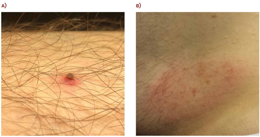
Bites from nymphal lone star ticks. A) uncomplicated bite in current study, and B) itchy Lyme disease like rash occurring after bite on the same author a year earlier. Reproduced under a Creative Commons Attribution License CC-BY 4.0 with the permission of J Goddard, JP Wyattt, https://doi.org/10.7759/cureus.29865.
New Insights
Of course, the straightforward recovery was good news for our dedicated researcher. But despite the frequency of human encounters with ticks, very little had previously been published on the progression of an uncomplicated tick bite, detailing the progress from the time of tick removal to lesion healing.
The medical assessment over 30 days, accompanied by photographic evidence, is likely the first account in the medical literature of the healing trajectory of an uncomplicated tick bite.
Dr Goddard and Dr Wyatt detail how small amounts of localised bleeding ensued following the initial bite of the tick and insertion of the hypostome. Skin irritation was greatest for the first four days, after which it subsided progressively over the 30 days. As the tick on Dr Goddard was removed promptly, the researchers argue that the development of the lesion in the following days was due to the saliva of the tick encountered at the initial bite.
The important message from this fascinating case study is that, even without complications, ticks may create skin lesions that are observable for at least 30 days. We should note that if a tick is not removed, as ensured in Dr Goddard’s case, it will continue to feed on the blood for several days (and, in doing so, rapidly increase in size). The resolution trajectories from tick bites in more complicated scenarios require further examination.
SHARE
DOWNLOAD E-BOOK
REFERENCE
https://doi.org/10.33548/SCIENTIA1015
MEET THE RESEARCHER

Dr Jerome Goddard
Mississippi State University
Mississippi State, MS
USA
Dr Jerome Goddard obtained his PhD in Medical Entomology from Mississippi State University in 1984. Directly afterwards, he served in the United States Air Force as a Captain and Medical Entomologist. In 1989, Dr Goddard was appointed State Medical Entomologist for the Mississippi State Department of Health, and in 2008, he took up his current position of Extension Professor at Mississippi State University. Dr Goddard has published over 200 scientific papers and 14 books. One of his medical textbooks, The Goddard Guide to Arthropods of Medical Importance, is in its seventh edition and was awarded ‘Highly Commended’ in the Public Health Category of the British Medical Association’s Best Medical Book of the Year in 2013. Dr Goddard has been a valuable asset to the entomological community throughout his esteemed career, as evidenced by his research, teaching and expert consultation services.
CONTACT
jgoddard@entomology.msstate.edu
W: https://www.biochemistry.msstate.edu/associate.php?id=10
KEY COLLABORATORS
Dr Julie P Wyatt, MD, Wyatt Dermatology Clinic
FURTHER READING
J Goddard, JP Wyatt, The Evolution of a Tick Bite Lesion, Cureus, 2023, 14(10), e29865. DOI: https://doi.org/10.7759/cureus.29865

REPUBLISH OUR ARTICLES
We encourage all formats of sharing and republishing of our articles. Whether you want to host on your website, publication or blog, we welcome this. Find out more
Creative Commons Licence (CC BY 4.0)
This work is licensed under a Creative Commons Attribution 4.0 International License. 
What does this mean?
Share: You can copy and redistribute the material in any medium or format
Adapt: You can change, and build upon the material for any purpose, even commercially.
Credit: You must give appropriate credit, provide a link to the license, and indicate if changes were made.
SUBSCRIBE NOW
Follow Us
MORE ARTICLES YOU MAY LIKE

Assoc Prof. Nicholas Brown | Rethinking Prostate Care: A New Frontier in Treating Benign Prostatic Hyperplasia
For millions of men, ageing brings with it a set of frustrating and often disruptive urinary symptoms. These symptoms, caused by benign prostatic hyperplasia, or BPH, can affect sleep, confidence, and overall quality of life. Traditionally, treatment follows a familiar path. Patients begin with medications, often for years, and may eventually progress to surgery if symptoms worsen. Yet this pathway is not without its drawbacks. Medications can cause side effects, while surgery carries risks and recovery time. In recent years, a minimally invasive interventional radiology procedure called prostate artery embolisation, or PAE, has begun to challenge this traditional model. At the forefront of this shift is a collaborative research group, led by Dr. Nicholas Brown of the University of Queensland, whose series of P-EASY studies has explored whether PAE could transform how BPH is treated, particularly at earlier stages.

Jean Lycke | Addressing Unmet Medical Needs in Mucosal Disease: A Close-to-Market Innovation Approach
Recurrent Aphthous Stomatitis (RAS) is an oral condition characterized by one or several painful mucosal ulcers. RAS affects a large proportion of the population and has a point prevalence of approximately 2–3%, daily. The etiology remains unknown, and there is currently no curative treatment. Most patients experience recurring episodes over time, with each episode typically lasting up to a week. Here, we describe the development of a mucoadhesive patch which, when applied over a RAS ulcer, provides rapid pain relief. The patch is easy for patients to apply when symptoms begin and has the potential to be used as an over-the-counter product. The development of the Mucocort mucoadhesive patch is an example of a Close-to-Market innovation strategy that embraces simplicity within a complex healthcare system. By simplifying the product concept, the team has reduced the number of regulatory steps required before market approval. This MedTech/Pharma innovation model, known as the “4R” framework – Re-purposing, Re-formulation, Re-positioning, and Re-patenting – has guided the program from concept to commercialization. In addition to the biodegradable mucoadhesive patch developed for RAS ulcers, the team is extending the innovation concept to a mucoadhesive gel formulation for the prevention and treatment of chemotherapy-induced mucositis. This gel-based program is being commercialized separately through MucoShield.
The Translational Asian Agerelated Macular Degeneration Program Phase 2 (TAAP-2): Reimagining the Future of Vision Care
Age-related macular degeneration, often abbreviated as AMD, is one of the leading causes of vision loss among older adults worldwide. In Asia, where populations are ageing rapidly, its impact is particularly profound. For many, the disease quietly erodes central vision, making everyday activities such as reading, driving, and recognising faces increasingly difficult. Against this backdrop, the Translational Asian Age-related Macular Degeneration Programme, or TAAP for short, has emerged as a bold and ambitious effort to confront the disease headon. Now in its second phase, TAAP-2 represents a significant evolution in both scientific scope and clinical ambition.

Ms. Aikaterini Dritsoula | Looking Beyond Snoring: How Hidden Airway Problems Shape Children’s Sleep
For many parents, a child’s snoring may seem harmless, even endearing. Yet in some cases, it signals something more serious. Obstructive sleep apnoea is a condition in which a child’s breathing is repeatedly disrupted during sleep. These interruptions can affect growth, behaviour, and learning. Children with this condition may toss and turn at night, struggle to concentrate during the day, or show signs of hyperactivity and fatigue. Traditionally, enlarged tonsils and adenoids have been seen as the main culprits. Surgery to remove them has long been considered the standard treatment. However, research led by Consultant ENT Surgeon Ms. Aikaterini Dritsoula of The Leeds Teaching Hospitals NHS Trust invites us to look deeper. Her work suggests that the story is often more complex, especially in very young children.