
Dr Dawn Bowles | An Extremely Challenging Environment: Understanding the Molecular and Physiological Responses to Space Travel
Environmental stressors have an adverse impact on mammalian physiology, although biological systems are adept at evolving in response to regularly occurring stressors. However, the biological alterations resulting from less frequently encountered stressors are incompletely understood. Dr Dawn Bowles and her colleagues at Duke University Medical School are conducting experiments into the effects of the space environment on astronauts to further our understanding of the impacts of this extremely challenging environment.
Space: A Multitude of Complex Stressors
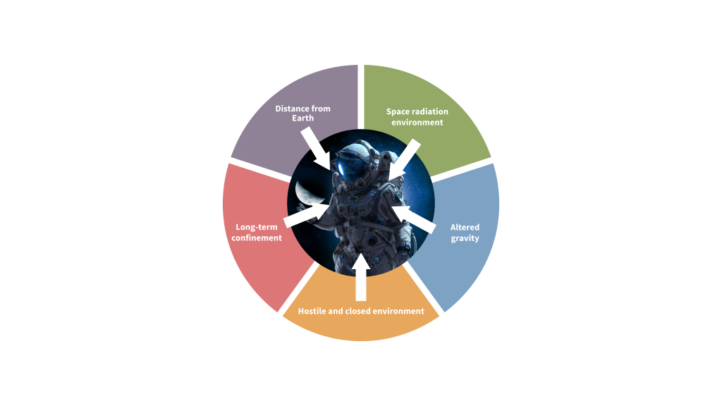
Astronauts, in their quest to explore and unravel the enigmatic entities that exist beyond Earth’s orbit, are increasingly confronting environments in which a wide range of complex environmental pressures, including microgravity and exposure to high levels of radiation, are par for the course.
To better understand the multitude of stressors experienced by astronauts, Dr Dawn Bowles and her colleagues in the Department of Surgery at Duke University Medical School, North Carolina, USA, have collaborated with a team at the National Aeronautics and Space Administration (NASA) to investigate the effect of such extreme environments on the perturbation of physiological systems.
The Price of Exploration
The pivotal role that gravity plays in the maintenance of physiological integrity is becoming increasingly apparent to scientists. While it is recognized that the absence of gravity is an extreme biological stressor, the exact impact that this has on the human body remains poorly defined. Given this, it follows that an environment with minimal gravity, so-called microgravity, may pose a high risk in terms of detriment to health. Likewise, exposure to chronic levels of high-energy radiation would logically pose significant health risks. Cellular stress responses in the body are varied and depend upon the type of stressor, cell type involved, number of cells affected, and the time since exposure.
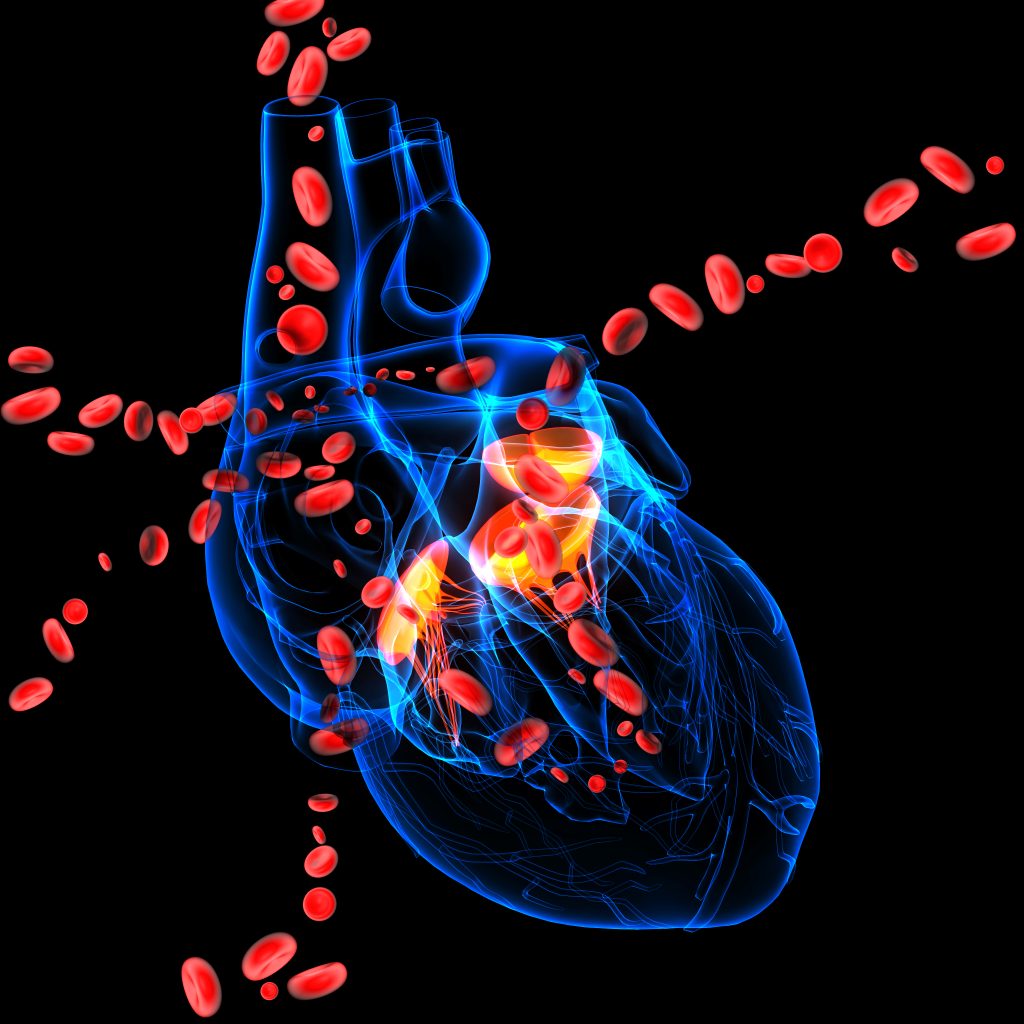
Astronauts returning from long-term space missions are susceptible to compromised immune function, decreased bone density, skeletal degeneration, and adverse cardiac effects including shrinkage of the heart muscle. These impacts all indicate that microgravity is a cellular stressor with the potential to adversely affect human health. This has clear implications for astronauts who are exposed to microgravity during space missions, and an understanding of how microgravity influences cellular molecular processes is imperative in comprehending these deleterious effects.
What we know so far about the pathways involved in the response to microgravity has been garnered from studies of astronauts and space flight. The predominant findings of such research include the alteration of cellular development, increased cell death and deterioration, modified protein turnover, and changes in the cytoskeleton (the structure that helps cells maintain their shape and internal organization, and helps them carry out vital functions). Although these biological amendments are known to be mediated by a complex system of genetic pathways, the precise proteins and mechanisms of action involved in the microgravity stress response have not yet been fully elucidated.
To determine the effects of microgravity on molecular processes within the cellular environment, Dr Bowles and her team employed a combination of well-established laboratory techniques to quantify large numbers of proteins, study the dynamics of alterations in protein content, and examine the biological pathways and networks involved in the response of heart cells to microgravity-induced stress.
Microgravity, Big Impact
Dr Bowles and her colleagues placed rat cardiac muscle cells into simulated microgravity or normal gravity conditions for a period of 12 hours, 48 hours, or 120 hours. The team reported that, while there were no differences or slight differences in the abundance of proteins after 12 and 48 hours in either condition, there was a noticeably higher difference in protein abundance after 120 hours of simulated microgravity. The team then proceeded to determine the protein turnover rate in cardiac muscle by using a novel modification of an established cell culture technique and measuring the incorporation of labelled amino acids into newly synthesized proteins. The results showed that there was a drastic reduction in protein turnover in the microgravity environment in comparison to the normal gravity environment over time.
Next, the team identified which proteins, biological processes, cellular components, and molecular functions were most significantly impacted by microgravity. Interestingly, those gene sets linked to the production and transport of energy in tissues with limited capacity for regeneration were up-regulated in both the protein turnover and abundance data sets. Conversely, the incidence of down-regulated gene sets was related to protein production and localization within the cell.
The team went on to investigate the mechanisms by which microgravity differentially influences cytoplasmic and structural processes. The researchers ruled out damage to the cell membrane, an increased rate of cell death, and increased protein degradation as potential causes of these differences in microgravity conditions compared to normal gravity conditions.
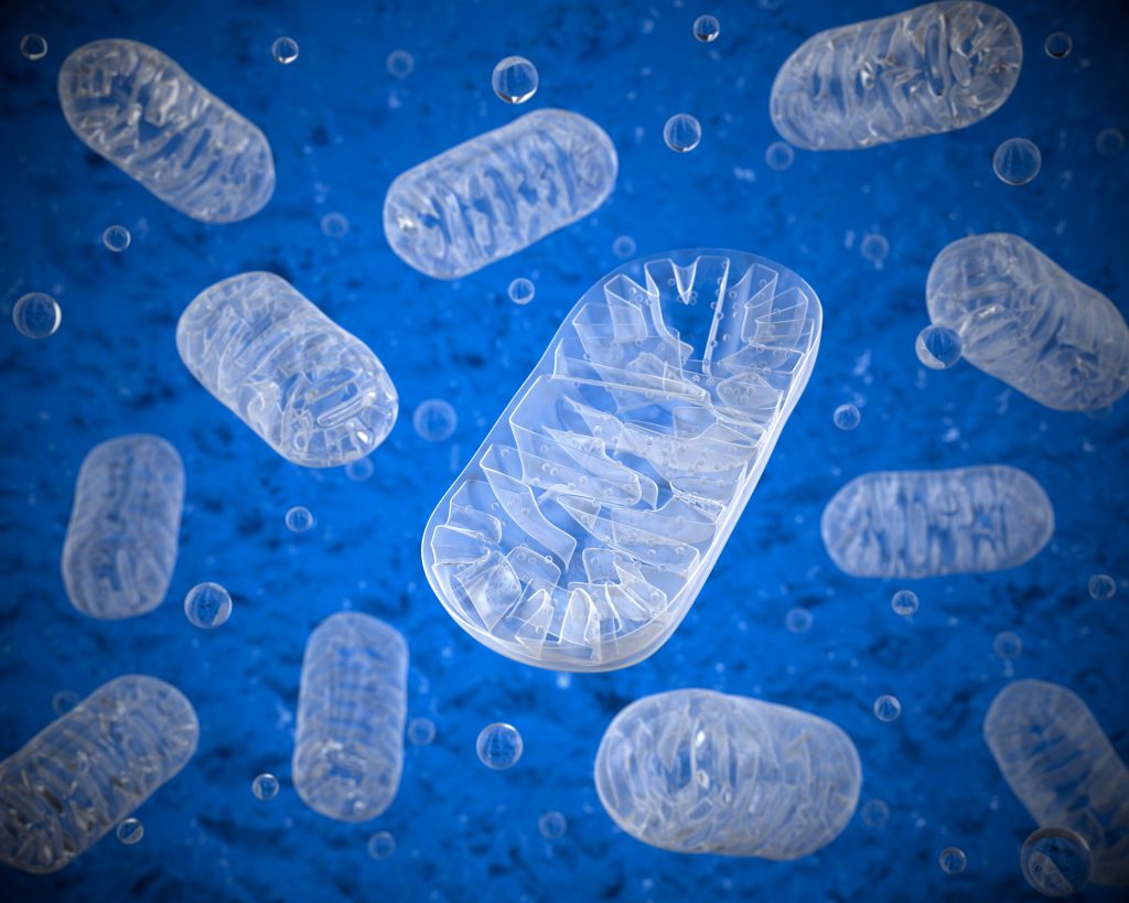
Taken together, these results suggest that the significantly diminished protein turnover and synthesis observed is due to a direct effect of exposure to microgravity for an extended time. Furthermore, their findings indicated that the integrity of the mitochondria, the so-called energy-producing powerhouse of the cell which comprises >30% of cardiac cell volume, is retained within cardiac muscle cells in the presence of microgravitational stress. This suggests that the mitochondria of cardiac cells respond to microgravity by supporting mitochondrial protein maintenance via a mechanism that confers preferential diversion of resources towards the mitochondria to preserve global cellular viability and promote short-term survival.
Previous findings have determined that, under favorable conditions, the specialized non-dividing cardiac cells known as cardiomyocytes maintain function by directing energy towards the maintenance of the contractile machinery and mitochondria by balancing protein synthesis and degradation.
Galactic Cosmic Rays
A unique component of the complex outer space environment is the high level of radiation comprised of Galactic Cosmic Rays (GCR). Studies involving survivors of acute high-level radiation exposure, such as nuclear accidents, have demonstrated that this is associated with a higher rate of delayed cardiovascular-related disease and death, although none of the published evidence relates specifically to space radiation. Whilst the Earth and its inhabitants are usually protected from radiation by the atmosphere, it is difficult to shield astronauts during long-term space missions, and thus they become more susceptible to serious cardiovascular health risks.
Indeed, a recent NASA study involving twins residing in space versus on Earth reported that genetic modifications and evidence of increased cardiovascular anomalies occurred only in the space-dwelling twin. However, whether this was attributable exclusively to radiation exposure remains unknown.
Accurately modelling the impact of radiation on the cardiovascular system has historically proven extremely challenging. But now, advances in engineering and physics technology are paving the way for a more authentic simulation of the space radiation environment. Taking advantage of this novel technology, Dr Bowles and her collaborators sought to determine the possible effects of GCR on the heart. Male mice of an age equivalent to a 34-year-old human being were exposed to a single dose of GCR and monitored over the course of one year for alterations in cardiovascular function using screening processes which mimicked routine assessment conditions. The team discovered that cardiac structure and function in GCR-exposed subjects were significantly altered when compared to non-irradiated controls or those exposed to other high-energy particles, although the physiological relevance of these changes was unclear.
The performance of the heart (cardiac contractile function) was assessed in mice one year post-exposure, and Dr Bowles and her team reported that those exposed to GCR demonstrated significantly lower function, increased vascular resistance, and increased arterial stiffness than the control subjects. Rather interestingly, despite these findings, GCR exposure did not confer an increase in mortality rates, blood pressure, or weight compared to controls when assessed at the one year mark. However, histological evaluation of cardiac tissues from GCR-exposed mice revealed increased cell degeneration, fragmentation, death, and a distorted cellular structure, suggesting long-term damage to the tissues of the heart following exposure to high-level radiation, which appears similar to that commonly seen in human congestive heart failure.
Dr Bowles has recently provided new molecular data indicating that mitochondria may be perturbed in these mice. This raises the question of what happens when you look at both effects simultaneously. Now, Dr Bowles are her team are doing exactly that – examining the combined effects of space radiation and microgravity.

The Mouse PAD
Alongside this work, in 2020 Dr Bowles and her colleagues described their establishment of a repository of over 5,000 tissues from mice exposed to space radiation. Importantly, the mouse Processing Aid Device (Mouse PAD) streamlines the preparation of materials, tissue collection, and tissue processing for research purposes. As stated by the researchers, the Mouse PAD is affordable, easy to use and produces high-quality, consistent results. It can also be easily adapted by other scientists who wish to establish their own unique biorepositories for research.
The Final Frontier
Dr Bowles and her colleagues are driving forward research aiming to explore the final frontier. Her fascinating studies have greatly contributed to the fundamental understanding of the response of heart cells to microgravity stress by using a novel application of an established laboratory method to successfully identify protein turnover rates whilst simultaneously monitoring protein expression changes within the cells. Furthermore, Dr Bowles and her team have demonstrated how global cellular protein turnover and synthesis is significantly reduced in cardiac cells exposed to microgravity, which is unrelated to increased cell damage, protein degradation, or cell death, and which may explain the mechanism via which heart muscle shrinkage in astronauts occurs. Moreover, the discovery that microgravity affects the entire cell equally and at the same time suggests that this particular stressor may induce an as-yet unreported cellular stress response.
The superbly executed research led by Dr Bowles also resulted in the discovery that astronauts exposed to GCR may suffer damage to vascular and cardiac tissues long after exposure, which may predispose them to conditions such as congestive heart failure.
Further studies to improve our understanding of the coordinated cellular response, the pathways involved in the body’s response, and physiological alterations emerging in response to stressors unique to the space environment will undoubtedly aid in the preparation of astronauts prior to extensive and far-reaching space missions. Work is already underway in Dr Bowles’ laboratory, showing an increasingly broad and holistic approach towards understanding the complex stressors experienced by astronauts.
This ground-breaking research will increase our understanding of the adverse biological effects of space travel and aid in developing risk models to ensure improved safety during and after explorations of life beyond our aerosphere.
SHARE
DOWNLOAD E-BOOK
REFERENCE
https://doi.org/10.33548/SCIENTIA904
MEET THE RESEARCHER
Duke Department of Surgery
Duke University Medical Center
Durham
North Carolina, NC
USA
Dr Dawn Bowles completed her PhD in Microbiology at Louisiana State University in 1999. After completing a postdoctoral position at the University of North Carolina, she took up several roles at Duke University Medical Center, where she is currently Assistant Professor in Surgery. Her extensive research spans the broad fields of medical genetics, cardiology, microbiology, molecular biology, surgery, and virology, and she has published prolifically in these fields with almost 100 peer-reviewed papers to date. She is the recipient of significant funding from prestigious award bodies, including the National Institutes of Health and NASA (National Aeronautics and Space Administration).
CONTACT
E: dawn.bowles@duke.edu
W: https://surgery.duke.edu/profile/dawn-elizabeth-bowles
KEY COLLABORATORS
https://www.bnl.gov/nsrl/
FUNDING
National Institutes of Health, USA
National Aeronautics and Space Administration, USA
University of Washington, USA
International Anesthesia Research Society
Baylor College of Medicine, USA
FURTHER READING
M Bishawi, FH Lee, DM Abraham, C Glass, et al., Late onset cardiovascular dysfunction in adult mice resulting from galactic cosmic ray exposure, iScience, 2022, 25(4), 104086. DOI: https://doi.org/10.1016/j.isci.2022.104086
ZD Brown, M Bishawi, JN Roan, et al., The use of an inexpensive processing aid device (the Mouse PAD) to facilitate rodent tissue banking, BioTechniques, 2020, 69(1), 364–368. DOI: https://doi.org/10.2144/btn-2019-0069
BJ Feger, JW Thompson, LG Dubois, et al., Microgravity induces proteomics changes involved in endoplasmic reticulum stress and mitochondrial protection, Scientific Reports, 2016, 6, 34091. DOI: 10.1038/srep34091
![]()
REPUBLISH OUR ARTICLES
We encourage all formats of sharing and republishing of our articles. Whether you want to host on your website, publication or blog, we welcome this. Find out more
Creative Commons Licence (CC BY 4.0)
This work is licensed under a Creative Commons Attribution 4.0 International License. 
What does this mean?
Share: You can copy and redistribute the material in any medium or format
Adapt: You can change, and build upon the material for any purpose, even commercially.
Credit: You must give appropriate credit, provide a link to the license, and indicate if changes were made.
SUBSCRIBE NOW
Follow Us
MORE ARTICLES YOU MAY LIKE

Assoc Prof. Nicholas Brown | Rethinking Prostate Care: A New Frontier in Treating Benign Prostatic Hyperplasia
For millions of men, ageing brings with it a set of frustrating and often disruptive urinary symptoms. These symptoms, caused by benign prostatic hyperplasia, or BPH, can affect sleep, confidence, and overall quality of life. Traditionally, treatment follows a familiar path. Patients begin with medications, often for years, and may eventually progress to surgery if symptoms worsen. Yet this pathway is not without its drawbacks. Medications can cause side effects, while surgery carries risks and recovery time. In recent years, a minimally invasive interventional radiology procedure called prostate artery embolisation, or PAE, has begun to challenge this traditional model. At the forefront of this shift is a collaborative research group, led by Dr. Nicholas Brown of the University of Queensland, whose series of P-EASY studies has explored whether PAE could transform how BPH is treated, particularly at earlier stages.

Jean Lycke | Addressing Unmet Medical Needs in Mucosal Disease: A Close-to-Market Innovation Approach
Recurrent Aphthous Stomatitis (RAS) is an oral condition characterized by one or several painful mucosal ulcers. RAS affects a large proportion of the population and has a point prevalence of approximately 2–3%, daily. The etiology remains unknown, and there is currently no curative treatment. Most patients experience recurring episodes over time, with each episode typically lasting up to a week. Here, we describe the development of a mucoadhesive patch which, when applied over a RAS ulcer, provides rapid pain relief. The patch is easy for patients to apply when symptoms begin and has the potential to be used as an over-the-counter product. The development of the Mucocort mucoadhesive patch is an example of a Close-to-Market innovation strategy that embraces simplicity within a complex healthcare system. By simplifying the product concept, the team has reduced the number of regulatory steps required before market approval. This MedTech/Pharma innovation model, known as the “4R” framework – Re-purposing, Re-formulation, Re-positioning, and Re-patenting – has guided the program from concept to commercialization. In addition to the biodegradable mucoadhesive patch developed for RAS ulcers, the team is extending the innovation concept to a mucoadhesive gel formulation for the prevention and treatment of chemotherapy-induced mucositis. This gel-based program is being commercialized separately through MucoShield.
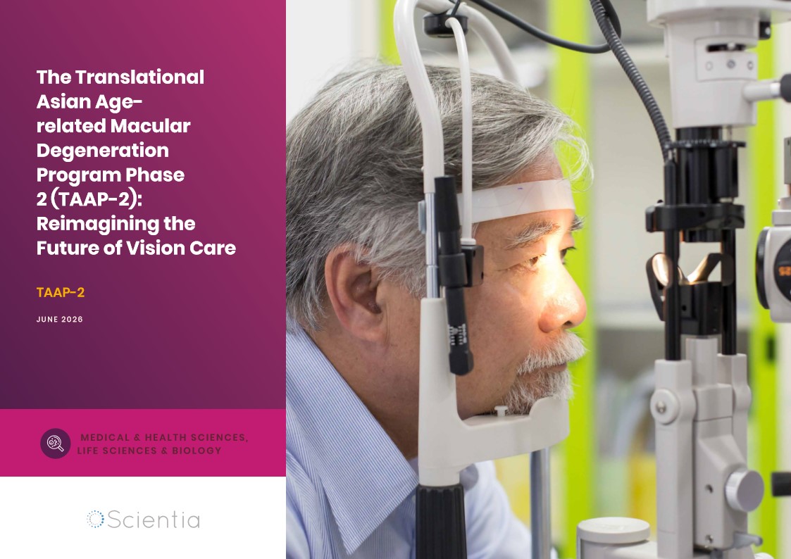
The Translational Asian Agerelated Macular Degeneration Program Phase 2 (TAAP-2): Reimagining the Future of Vision Care
Age-related macular degeneration, often abbreviated as AMD, is one of the leading causes of vision loss among older adults worldwide. In Asia, where populations are ageing rapidly, its impact is particularly profound. For many, the disease quietly erodes central vision, making everyday activities such as reading, driving, and recognising faces increasingly difficult. Against this backdrop, the Translational Asian Age-related Macular Degeneration Programme, or TAAP for short, has emerged as a bold and ambitious effort to confront the disease headon. Now in its second phase, TAAP-2 represents a significant evolution in both scientific scope and clinical ambition.
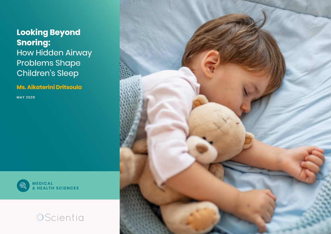
Ms. Aikaterini Dritsoula | Looking Beyond Snoring: How Hidden Airway Problems Shape Children’s Sleep
For many parents, a child’s snoring may seem harmless, even endearing. Yet in some cases, it signals something more serious. Obstructive sleep apnoea is a condition in which a child’s breathing is repeatedly disrupted during sleep. These interruptions can affect growth, behaviour, and learning. Children with this condition may toss and turn at night, struggle to concentrate during the day, or show signs of hyperactivity and fatigue. Traditionally, enlarged tonsils and adenoids have been seen as the main culprits. Surgery to remove them has long been considered the standard treatment. However, research led by Consultant ENT Surgeon Ms. Aikaterini Dritsoula of The Leeds Teaching Hospitals NHS Trust invites us to look deeper. Her work suggests that the story is often more complex, especially in very young children.